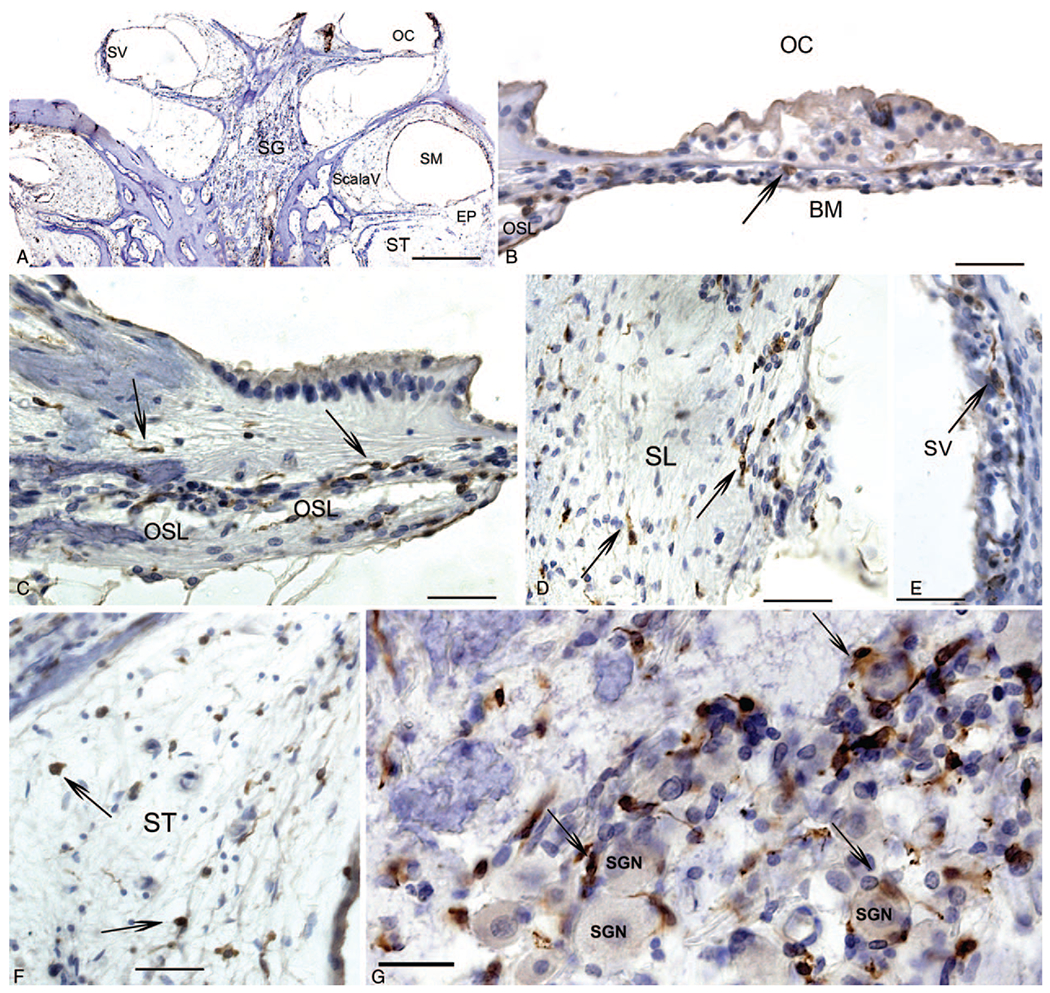
FIG. 4.

Immunohistochemistry for Iba1 macrophages in another CI cochlea section. A, Low magnification view to indicate the location of Iba1 IR macrophages in the cochlea: visualizing the stria vascularis (SV), the organ of Corti (OC), Scala Vestibuli (Scala V), the Scala Media (SM) which exhibits hydrops, and the Scala tympani (ST). B, Organ of Corti (OC), thin arrows point to Iba1-IR round macrophages, within the basilar membrane (BM). C, Iba1-IR elongated macrophages in the osseous spiral lamina (OSL). D, Iba1-IR macrophages in the spiral ligament (SL). E, Iba1-IR macrophages in the stria vascularis (SV). F, Iba1-IR macrophages within the areas of fibrosis within the scala tympani (ST). G, Iba1-IR macrophages in the spiral ganglia region, some surrounding spiral ganglia neurons (SGN) showing a ramified morphology with spider-like extensions (long thin arrows). Magnification bar in A = 500 μm, in B to F = 40 μm. CI indicates cochlear implant; Iba1, ionized calcium binding adaptor 1; IR, immunoreactive.
