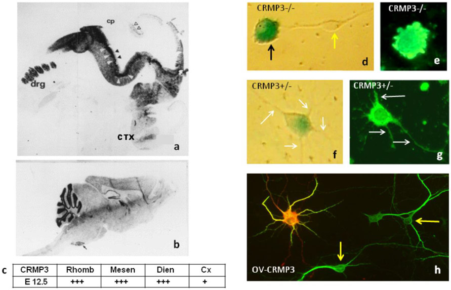
Figure 1. Expression of CRMP3/DPYSL4 during nervous system development and role in cultured hippocampal neurons.
In situ hybridization of CRMP3 in sagittal sections of embryonic and adult mouse brain. (a) At E12.5, transcripts were detected in dorsal root ganglia (drg) and the central nervous system (CNS). Different regions of the rhombencephalon (Rhomb), mesencephalon (Mesen), and diencephalon (Dien) were stained and are denoted with white arrows. The black arrows denoted the CRMP3 expression in the ventricular neuroepithelia and the cortex (Cx). Staining was also observed in cerebellum, which is marked with unfilled arrows. No staining was observed in the choroid plexus (cp). (b). In sections obtained from adult mice, CRMP3 mRNA was detectable mainly in the cerebellar granular layer, the inferior olive, and the dentate gyrus. (c). Summary of In Situ Hybridization with an antisense riboprobe: highly positive signal (+++); weak signal (+). Representative Crmp3−/−cells with no visible processes (d; e) and Crmp3+/− cell with neurites (f; g) using β-galactosidase activity (d; f) or immunostaining with MAP2 antibodies (e; g). Transfected with Fl-CRMP3, neurons are characterized by active transport of the protein, increased formation of lamellipodia, and increased dendritic arborization. Representative CRMP3-over-expressing neuron (h; ov-crmp3) immunostained for Flag (red) and the dendritic marker MAP2 (green). Overlay of image from transfected neurons are in yellow (h, right). Yellow arrows show control (h) or LacZ− (d) cells. White arrows show neurites (f, g).

