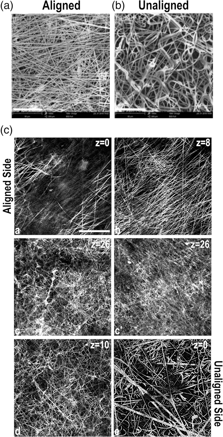
FIGURE 1.

Fiber orientations in a multilayered electrospun scaffold. Representative scanning electron microscope (SEM) images of the aligned (a) and unaligned (b) sides of the E1001(1k) scaffold are shown. Scale bar = 80 μm. (c) Confocal microscopy images were collected in reflectance mode to capture the structure of the scaffold fibers. Then, 2 μm thick confocal slices are shown starting from aligned (images a–c) and unaligned (images c′–e) sides. z corresponds to the distance from the scaffold surface. Images are representative of scaffold structure. Scale bar = 50 μm
