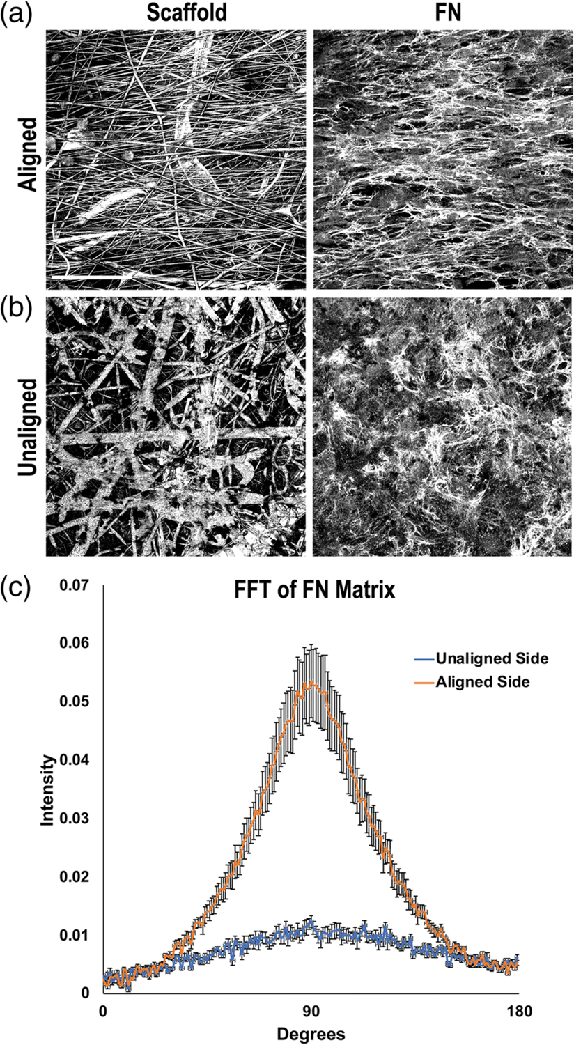
FIGURE 2.

Fibronectin (FN) matrix fibril orientations on the electrospun scaffold. Mouse NIH 3T3 fibroblasts were seeded onto either the aligned (a) or unaligned (b) side of separate scaffolds and grown for 7 days. Cells were fixed and stained with R457 anti-FN antiserum. Representative reflectance (scaffold) and FN confocal images of the same fields of view are shown. Scale bar = 50 μm. (c) FN fibril orientations on either side of the scaffold were quantified by fast Fourier transform (FFT) analysis of fluorescent images. Five images were averaged per condition and radial intensity data were plotted from 0 to 180° (error bars = SEM)
