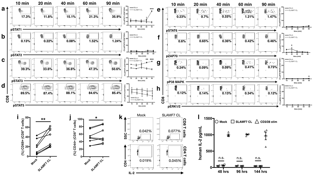
Figure 4. Alterations in T cell signaling pathways following SLAMF7 activation.

(a-h) Time course of isolated primary human CD8+ T cells stimulated in vitro by SLAMF7 cross-linking (n=2 donors). Representative biaxial plots are shown for only SLAMF7 CL conditions. Surface expression of CD69 (i) and CD44 (j) was assessed on (n=8) donors following 6 days of in vitro stimulation. (k) Biaxial plots showing lack of IL-2 expression in CD8+ (top) and CD4+ primary human T cells following 3 days of in vitro stimulation (n=2), representative of two independent experiments. (l) Secretion of IL-2 by in vitro stimulated primary human CD3+ T cells was assessed over time by ELISA (n=4). No exogenous IL-2 was added to cultures and results are representative of two independent experiments. (a-h) is representative of two independent experiments with a total of 5 healthy donors and was analyzed by two-way ANOVA with Sidak’s multiple comparison test. (i, j) contains pooled samples from 2 independent experiments and was analyzed with a paired students t-test. Two-way ANOVA with Tukey’s multiple comparison test used for (l)
