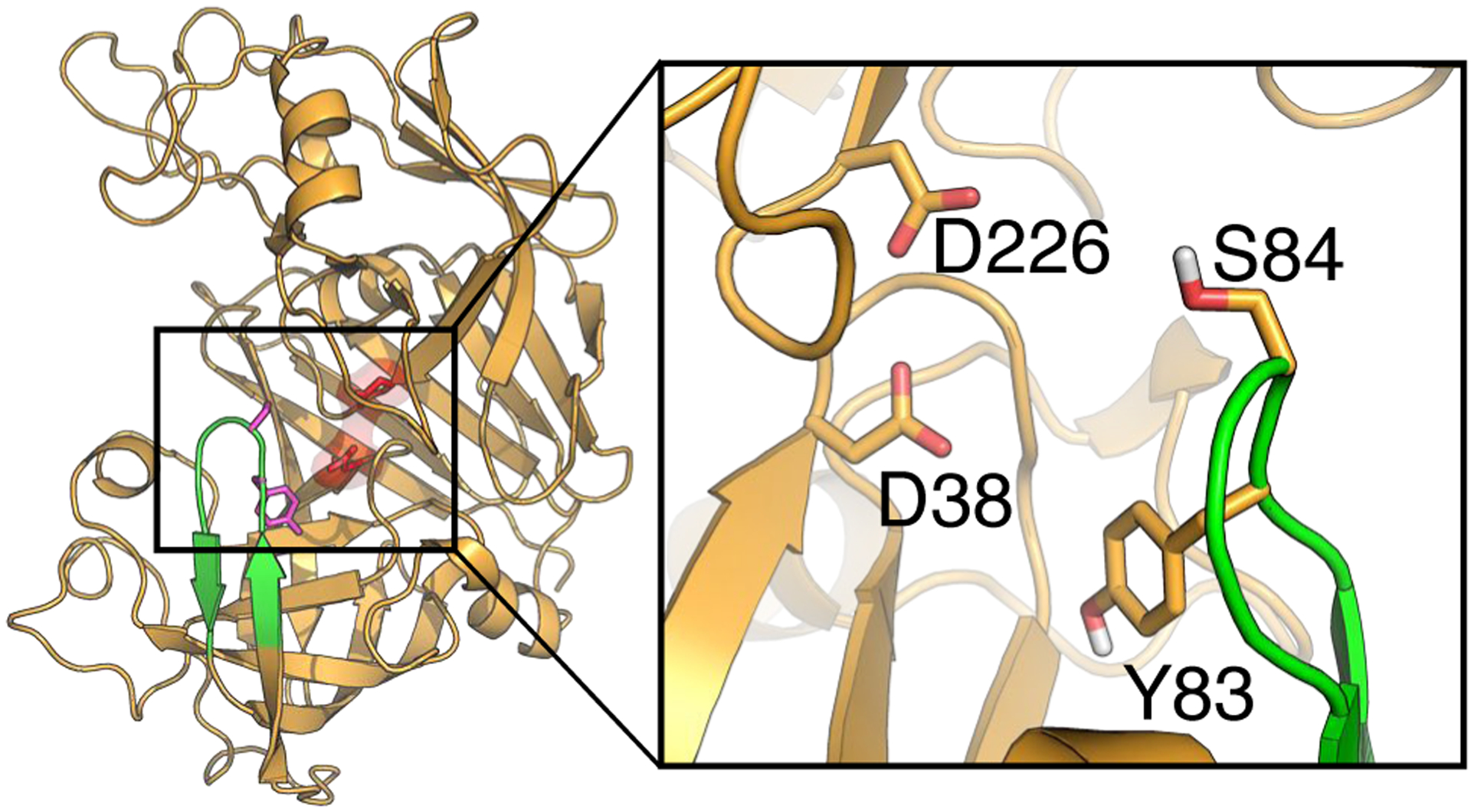
Figure 2. The overall structure of human renin and its substrate binding site.

The X-ray crystal structure of human renin (PDB: 2ren14), with the flap (residues Thr80 to Gly90) and dyad (Asp38 and Asp226, in surface rendering) colored green and red, respectively. A zoomed-in view of the substrate binding site and flap region is shown. Residues discussed in the main text are labeled.
