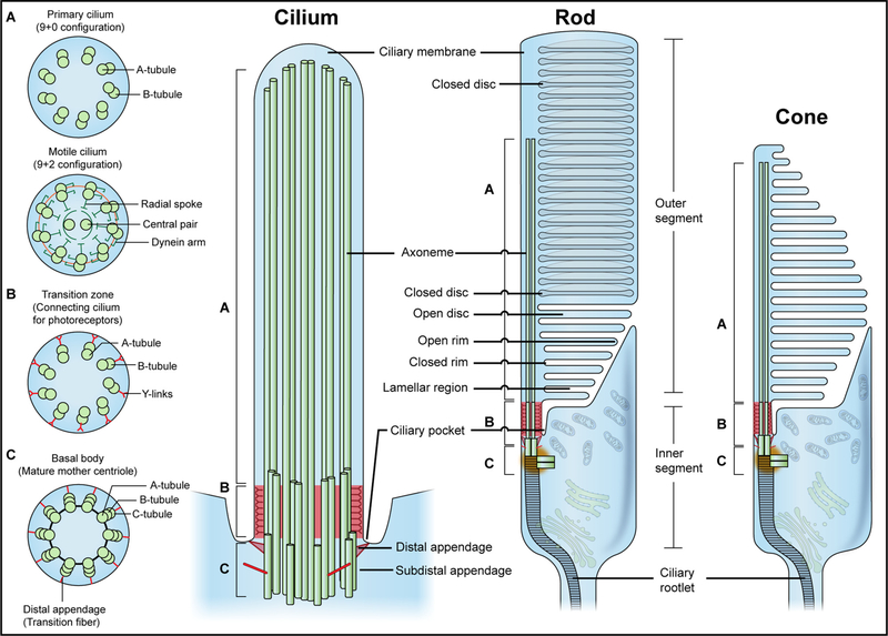
Fig. 1.
The architecture of the general primary cilium (left) and the photoreceptor outer segment (right). The primary cilium consists of a ciliary membrane and an axoneme. The ciliary membrane is continuous with the plasma membrane but differs in compositions to regulate diverse signaling pathways. In most mammalian cells, the plasma membrane invaginates at the base of the axoneme, forming the ciliary pocket for endocytosis and docking of intraflagellar transport particles. The axoneme elongates from the basal body (BB), which is the mature mother centriole with distal appendages and subdistal appendages. Cross-section diagrams at different positions of the primary cilium with a doublet (axoneme) (“9 + 0” configuration), doublet with Y-links (transition zone), and triplet microtubule structure (basal body) are shown in upper A, B, and C insets, respectively. In motile cilia, the axoneme displays “9 + 2” configuration, with an additional pair of microtubules in the center (Lower A inset). Photoreceptors feature a gradual doublet (base) to singlet (tip) microtubule transformation in the ciliary axoneme [125]. Photoreceptors harbor distinct features to accommodate their sensory function: the outer segment contains tightly packed discs with phototransduction machineries to efficiently capture photons; the inner segment possesses the endoplasmic reticulum, Golgi apparatus, and a large number of mitochondria to meet the high energy demand and biosynthetic needs of the photoreceptors; the ciliary rootlet anchors the BB to the inner segment to stabilize the outer segment.

