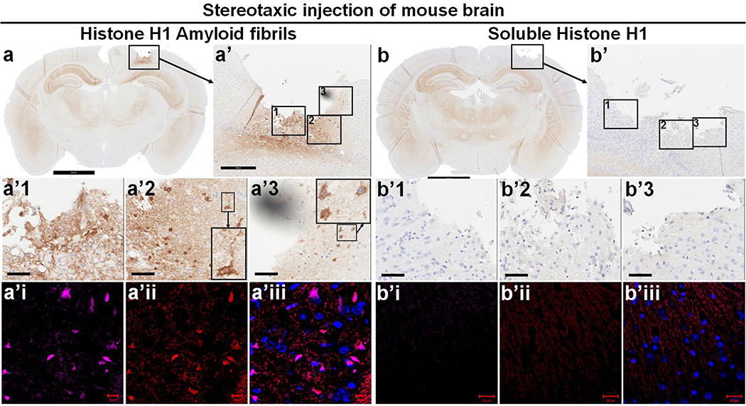
Fig. 4. Amyloid fibrillar, but not soluble histone H1, can induce αS aggregation in vivo upon inoculation into mouse brain.
Soluble recombinant human histone H1 and derived histone amyloid fibrils were inoculated into somatosensory cortex of wild-type FVB mice at 10 months of age via stereotaxic injection. Brains were harvested a week later. Paraffin sections of brain injected with amyloid fibrillar histone H1 (a) or soluble histone H1 (b) were subjected to immunohistochemical staining with antibody to αS (610787, BD Biosciences) to demonstrate αS pathology. Scale bar 2 mm. Panels a’ and b’ show enlarges framed areas in a and b. Scale bar 300 μm. The framed areas in a’ and b’ were further enlarged to a’1, a’2, a’3, b’1, b’2 and b’3 to show that amyloid histone H1 fibrils could induce intracellular αS deposits and thread- or dot-like αS pathology in the brain. Brain regions corresponding to a’ and b’ in adjacent sections were subjected to immunofluorescence staining with primary antibodies to human histone H1 (ab125027, Abcam, a’i & b’i) and αS (610787, BD Biosciences, a’ii & b’ii) with goat secondary antibodies to rabbit (Alexa Fluor 647) and mouse (Alexa Fluor 568). Scale bar 10 μm. Note: There are two inset pictures in both a’2 and a’3 in which the picture of small size is enlarged aside.

