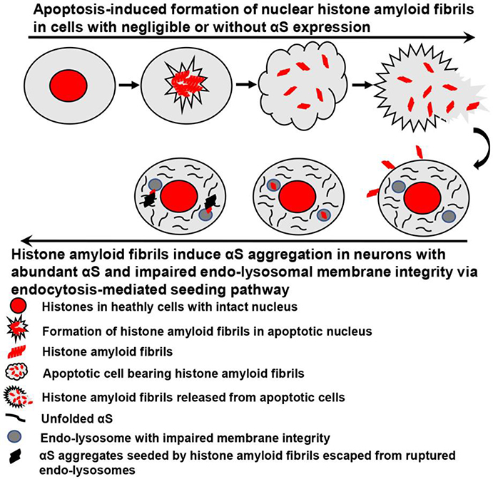
Fig. 5. The formation and spreading of apoptosis-induced histone amyloid fibrils and subsequent induction of αS aggregation in recipient neurons.
This schematic diagram only focuses on formation of nuclear histone amyloid fibrils in cells with negligible or without αS expression. For apoptotic cells bearing abundant αS, histones can directly interact with αS to rapidly form αS aggregates, leading to the propagation of αS pathology in surrounding cells, which was discussed in our previous studies[10, 12].

