
Figure 1.
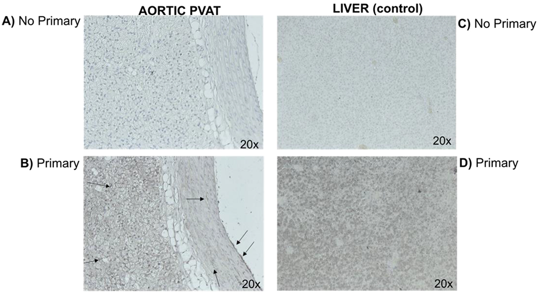
Immunohistochemical staining of Flavin-Mono-Oxidase-3 (FMO3) in the rat thoracic aorta (left) and rat liver (right) as a positive control for FMO3 expression. Section incubated without (panels A and C) and with primary FMO3-specific antibody (B and D). Images (magnification 20x) depict representative of a minimum of four separate animals. Adjustments in brightness and contrast were made to the whole panel of a photograph, not a portion. Arrows indicate the presence of staining for FMO3. Representative of five (5) separate animals.