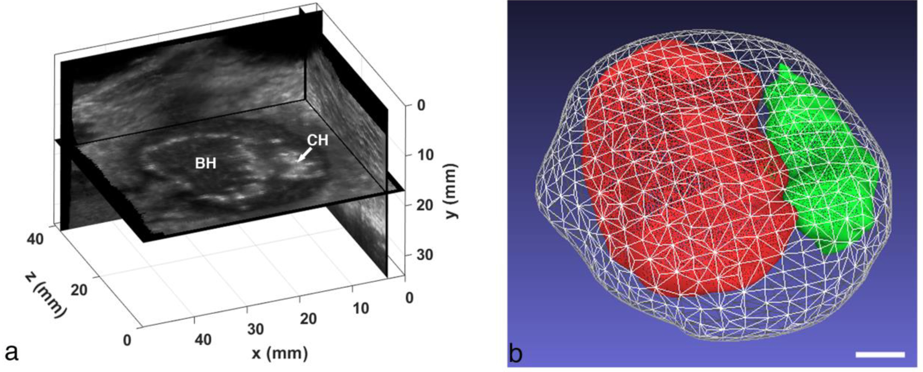
Figure 6.

(a) Orthogonal views from a 3D volume reconstruction of the ex vivo abscess that was treated with BH and CH. The “constant depth” image plane showing the BH and CH regions is unique to the reconstruction. (b) Surface reconstructions of the full abscess (white mesh), BH treatment region (red) and CH region (green). The total abscess volume is 14.6 cc (BH volume: 4.9 cc, CH volume: 1.2 cc). Scale bar represents 1 cm.
