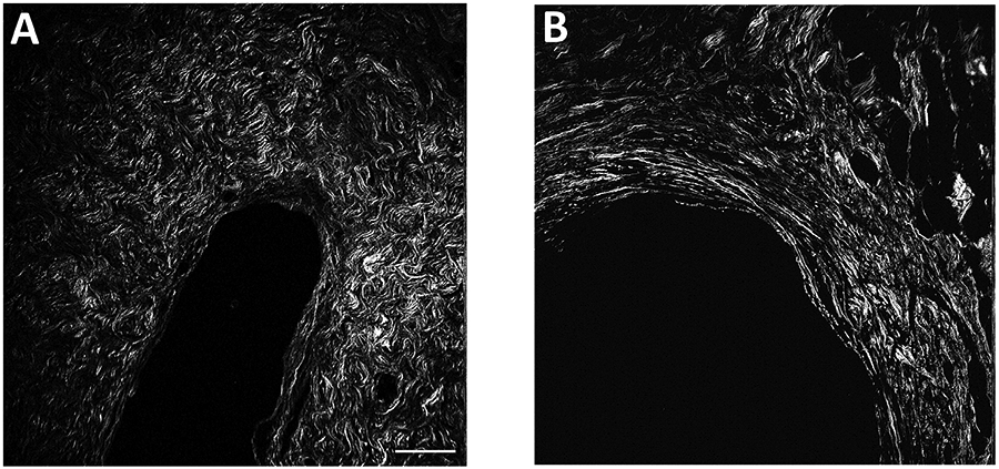
Figure 1.

Sample multiphoton microscopy images of collagen fibers, illustrating cases with different collagen features. Panel A shows an area of DCIS surrounded by short, curvy, dense fibers. Panel B shows an area of DCIS surrounded with long, straight, high alignment fibers. Scale bar, 100 μm.
