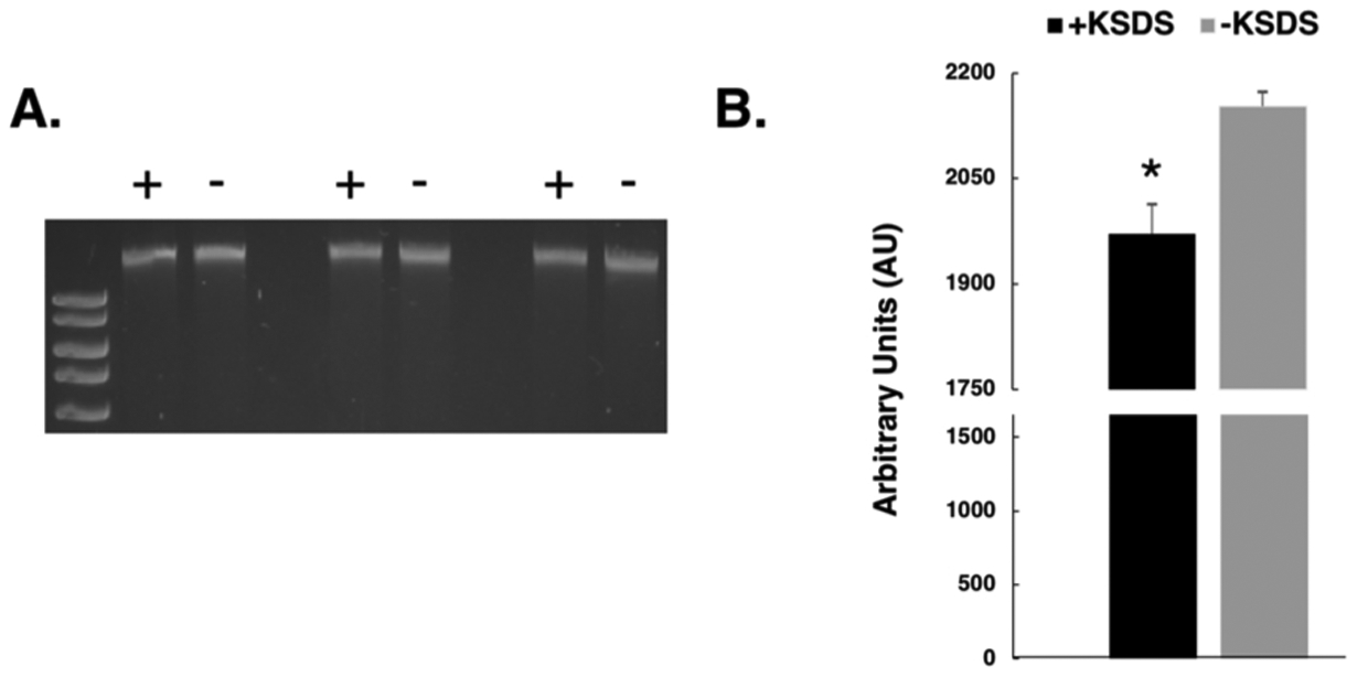
Fig. 5. Formation of cisplatin-induced DNA-protein crosslinks in human cells.

Human HT1080 cells were treated with 100μM cisplatin for three hours and mitochondrial DNA purified as described in methods. A. Purified DNA was divided in equal aliquots which were incubated in the presence (+) or absence (−) of KCl/SDS and DPC-containing DNA sedimented by centrifugation as described in the methods section. Soluble material was electrophoretically resolved on a 0.8 % agarose gel in triplicate and stained with ethidium bromide. B. Scanning densitometry was performed on the gel to determine the relative amount of mitochondrial DNA present in the KCl/SDS treated (black bar) and untreated samples (grey bar). Results depict the mean amount of DNA present +/− SEM, N = 3. *, P < 0.05, Student t-test.
