
Fig. 3.
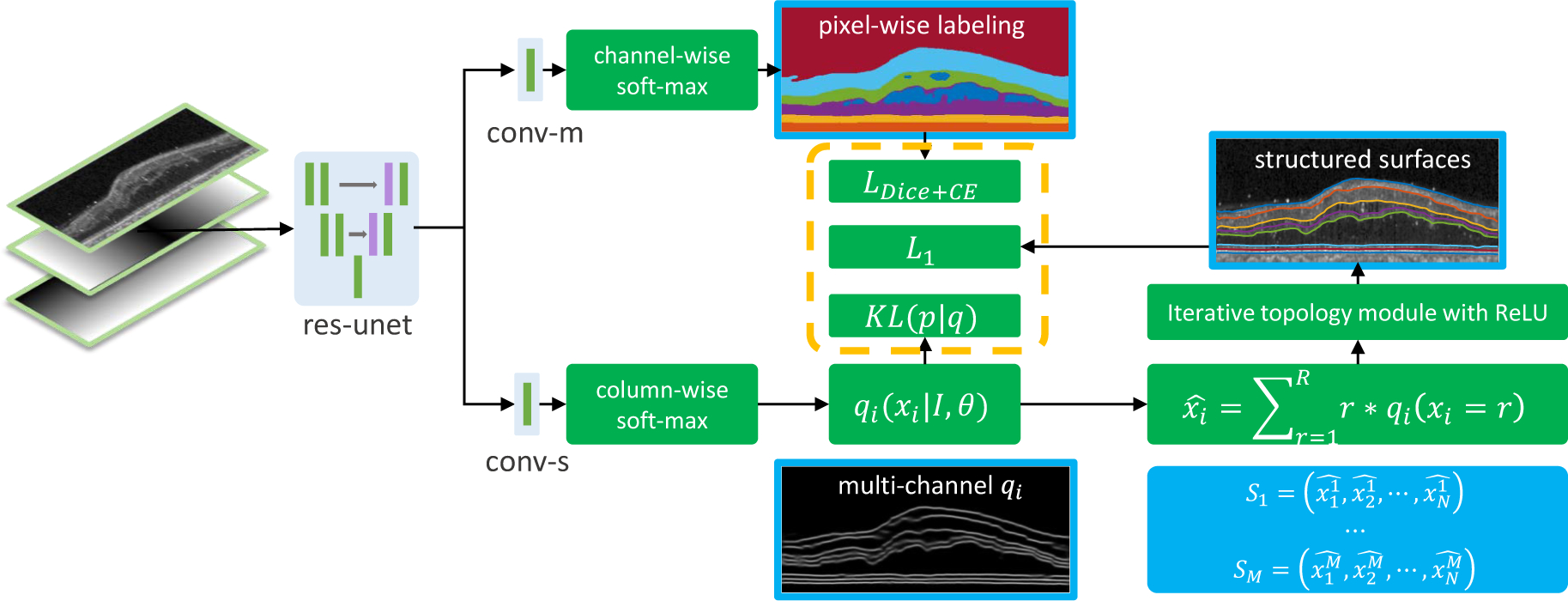
A schematic of the proposed method. The input is a three channel image (flattened B-scan, normalized x and y coordinates). The features extracted by the res-unet (a residual U-Net) are shared by the two output branches. The top branch (conv-m) uses channel-wise softmax to output segmentation probability maps for the layers, backgrounds (choroid or vitreous), and lesions. The bottom branch (conv-s) uses column-wise soft-max to produce variational qi’s and then uses soft-argmax and iterative ReLU to produce M structured surfaces.