Figure 2.

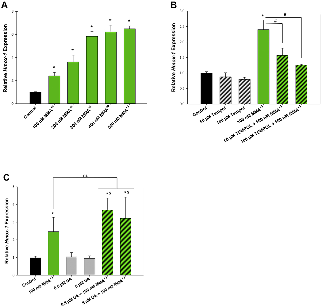
MMA+3, but not UA induces oxidative stress in mouse thymus cells. Mouse thymus cells were exposed in vitro to MMA+3 or UA for 4 h and Hmox-1 expression was measured by qPCR. (A) Dose dependent increase of Hmox-1 expression. (B) The antioxidant, Tempol significantly reverses MMA+3-induced Hmox-1 expression. (C) Exposure to UA at 0.5 and 5 μM does not induce Hmox-1 expression. Data are presented as mean ± SD. *Statistically significant difference (p<0.05) in one-way ANOVA followed by a Dunnett’s t-test compared to the control group. Panel A: one-way ANOVA followed by a Dunnett’s t-test; panels B and C: one-way ANOVA followed by a Tukey’s multiple comparisons post hoc test. *Statistically significant difference (p<0.05) compared to the control group. #Statistically significant difference (p<0.05) compared to 100 nM MMA+3. $Statistically significant difference (p<0.05) compared to individual UA doses, ns, no statistically significant difference compared to 100 nM MMA+3.
