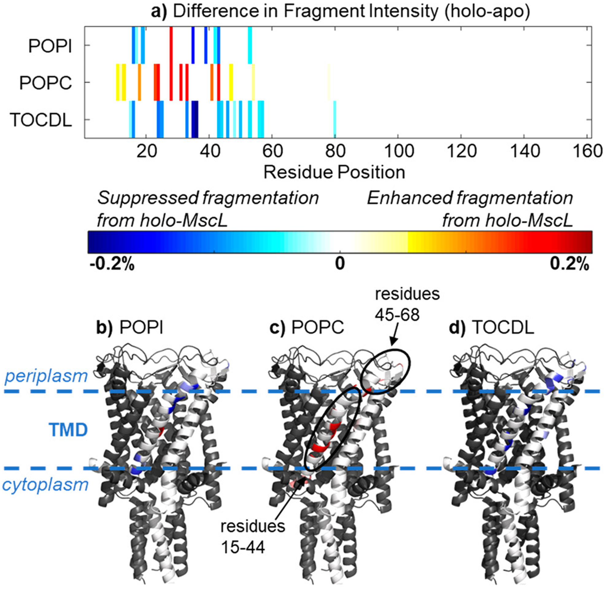
Figure 5.

Differences in the UVPD fragment intensity between apo-MscL and holo-MscL bound to five lipids plotted by (A) residue position and (B-D) on a subunit of the crystal structure. The circled regions showed the most significant variations between apo-MscL and holo-MscL. Reprinted from Sipe, S. N.; Patrick, J. W.; Laganowsky, A.; Brodbelt, J. S. Enhanced Characterization of Membrane Protein Complexes by Ultraviolet Photodissociation Mass Spectrometry. Anal. Chem. 2020, 92, 899–907 (ref 127). Copyright 2020 American Chemical Society.
