FIG. 2.
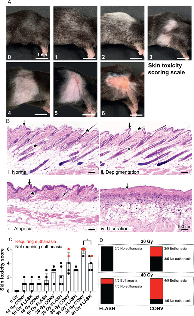
FLASH irradiation results in reduced severe skin toxicity compared to CONV dose-rate irradiation at high doses. Panel A: Skin toxicity scoring scale. Lesions were graded on a 0–6 scale using the following criteria:← 0 = normal; 1 = <50% depigmentation within the radiation field; 2 = ≥50% depigmentation within the radiation field; 3 = <50% alopecia (± depigmentation) within the radiation field; 4 = ≥50% alopecia (± depigmentation) within the radiation field; 5 = <50% ulceration (± alopecia and depigmentation) within the radiation field; 6 = ≥50% ulceration (± alopecia and depigmentation) within the radiation field. Representative images are shown of the scores assigned. Inter-rater reliability score (Cohen’s kappa) between two observers blinded to the interventions was 0.91. Scale bar = 1 cm. Panel B: Representative histologic images (H&E stained) of (i) normal mouse skin, (ii) depigmentation, (iii) alopecia and (iv) ulceration. In the normal mouse skin, the epidermis is 1–2 cell layers thick (i, black arrow) and there are numerous, evenly-spaced anagen hair follicles (i, asterisks) containing pigmented hair shafts. Mice exhibiting depigmentation retain the 1–2 cell layer thick epidermis (ii, black arrow) and evenly-spaced anagen hair follicles, but have increased numbers of depigmented hair shafts (ii, asterisks). Mice exhibiting alopecia have fewer, unevenly-spaced catagen hair follicles (iii, asterisks) and epidermal hyperplasia (iii, black arrow). Mice with ulceration have complete loss of the epidermis (iv, black arrow) and all associated hair follicle structures. Histologically, we observed no visible differences between FLASH and CONV irradiation in the character of these pathologic features except in frequency or severity grossly. Scale bar = 100 μm. Panel C: Severity of skin toxicity per cohort at 8 weeks postirradiation. The 40 Gy FLASH-irradiated cohort had significantly lower scores than the 40 Gy CONV-irradiated cohort; n = 5 per cohort; red dots indicate animals that had to be euthanized for meeting skin toxicity criteria; bars represent mean ± SD. *P < 0.05. Panel D: Mice in high-dose irradiated cohorts that met euthanasia criteria by week 8 postirradiation. Only mice in the 30 and 40 Gy CONV irradiated, and 40 Gy FLASH irradiated cohorts had to be euthanized for meeting skin toxicity criteria. Each bar represents a fraction of the total in each cohort that either met or did not meet euthanasia criteria. These results indicate a skin-sparing effect of FLASH compared to CONV irradiation at high doses of 30–40 Gy in a single fraction. At doses up to 20 Gy, there was no ulceration in either FLASH or CONV irradiated animals at 8 weeks.

