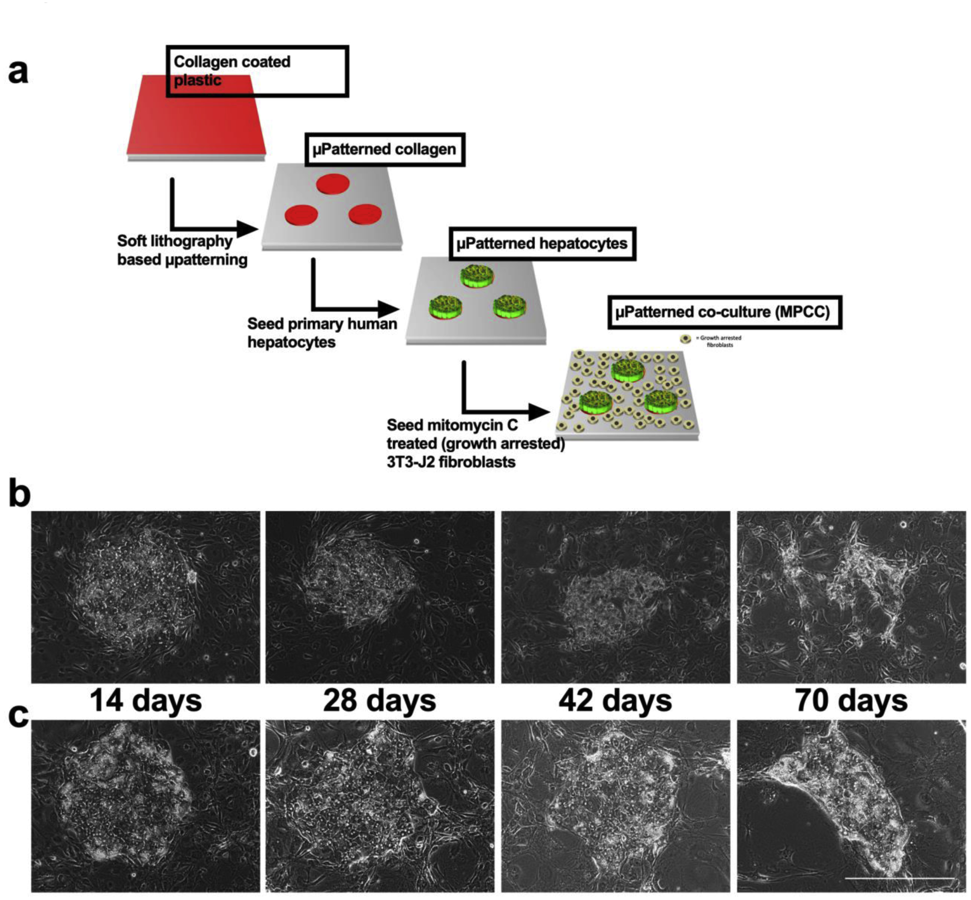
Figure 1. Schematic for MPCC fabrication and hepatocyte morphology over time.

(a) MPCC fabrication schematic. Representative phase contrast images over time of PHH islands surrounded by fibroblasts in MPCCs cultured in the (b) traditional medium and (c) physiologic medium. Scale bar represents 400 μm.
