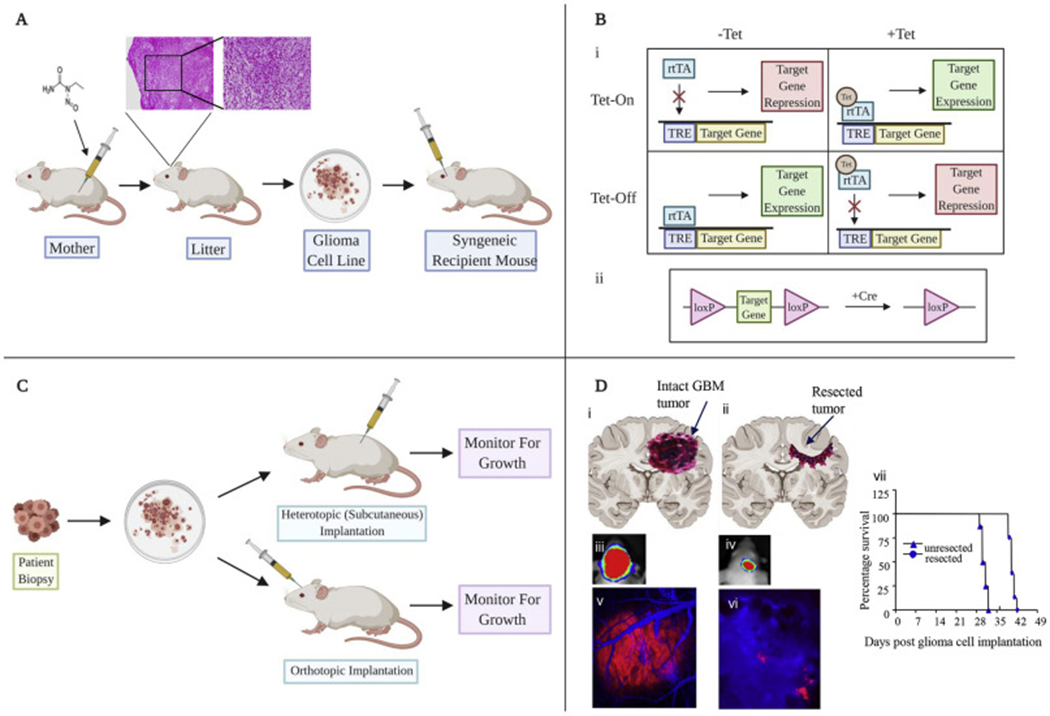
Figure 1: In vivo models of glioblastoma in rodents.

A) Traditional methods include ENU administration in pregnant rodents leading to tumor formation, which can be harvested and processed into cell lines in vitro. B) Genetically engineered systems include reversible systems using Tet regulation (i) or Cre recombinase (ii). C) Patient derived xenografts can be injected subcutaneously or directly into cerebral cortex. D) Resection models designed to recapitulate the tumor environment following primary resection of tumors. (i,ii) Cartoons showing GBM tumors before and after tumor resection in the brain mice. (iii-vii). Mice with established GBM-Fluc-mCherry GBMs were imaged by bioluminescence imaging (iii,iv) and intravital microscopy (v,vi) before and after tumor resection. Kaplan-Meier survival curves of mice with and without resected U87-Fluc-mCherry tumors (vii) (adapted from Kauer et al 2012) (82).
