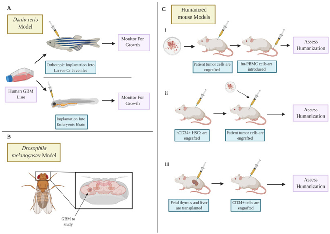
Figure 2: Alternative models of glioblastoma.

A) The zebrafish model using both embryos and adult species allow modeling of the disease to be performed quickly and can allow tumors to be imaged in real time. B) The drosophila model allows the study of glioblastoma using various genetic manipulations. C) Humanized mouse models allow modeling of tumors with partially humanized immune systems using three methods. (i) hu-PBMC cells are introduced after patient tumor cell engraftment. (ii) hCD34+ stem cells are engrafted before the patient tumor cells are added. (iii) Humanized mouse models can be created through the transplantation of fetal liver and thymus under the kidney capsule. CD34+ cells are engrafted afterwards.
