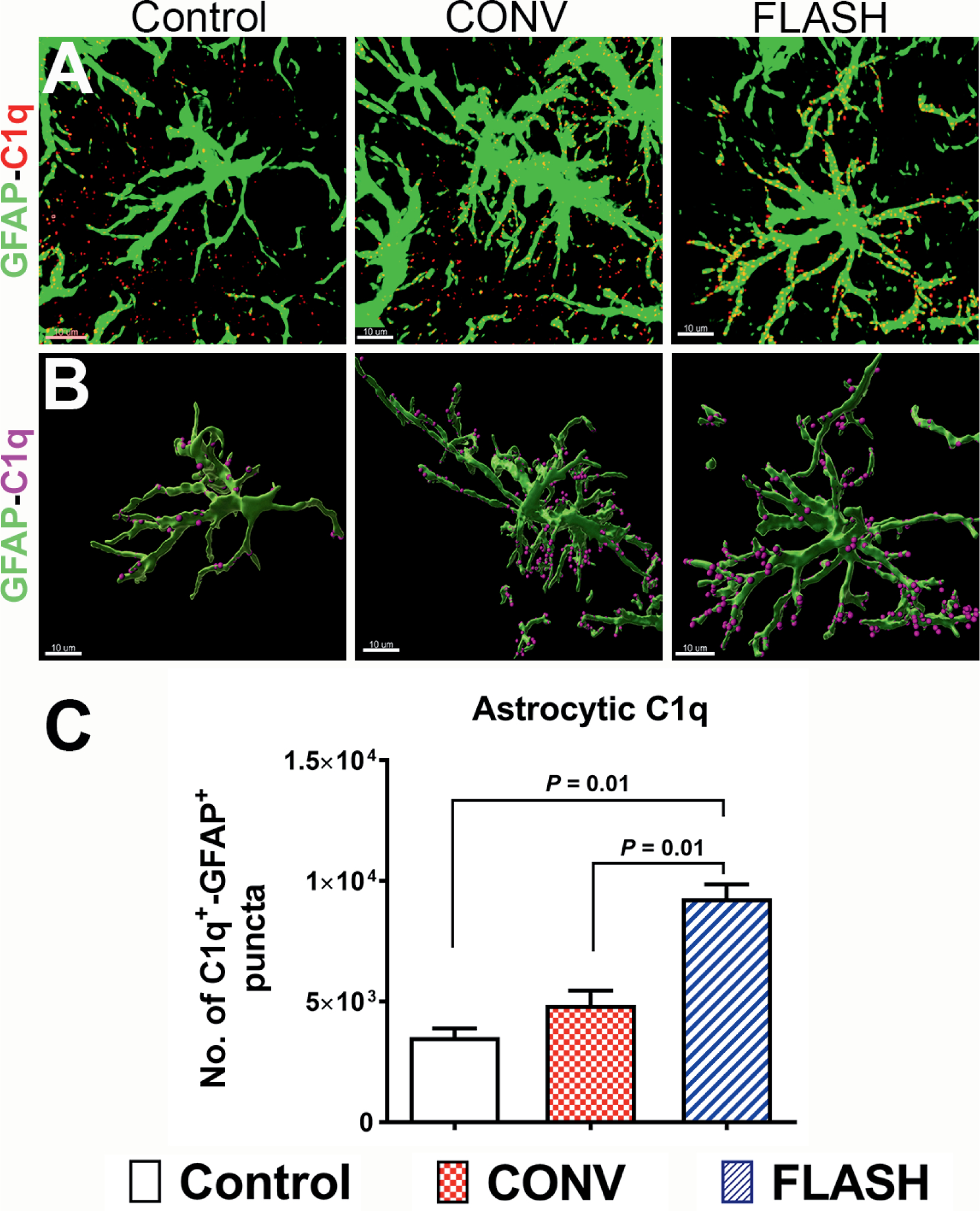
FIG. 3.

Elevated astrocytic expression of complement C1q in the irradiated brain. Volumetric quantification of confocal z stacks (green, GFAP; red, C1q, panel A) and 3D reconstruction of GFAP+ astrocytes (green, panel B) co-labeled with C1q (magenta spots, panel B) showed a significantly elevated complement C1q one month after either 10 Gy CONV-RT or FLASH-RT (panel C). Data are presented as mean ± SEM (n = 4–6 animals per group). P values are derived from non-parametric Kruskal-Wallis H test and Mann-Whitney’s comparison between each group as indicated. Scale bar = 10 μm (panels A and B).
