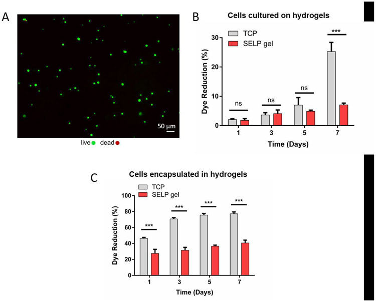
Fig. 5. Cell viability.
Cytocompatibility test of L929 murine fibroblasts grown on or encapsulated within the SELP-CarHc hydrogels. A. Live/dead fluorescent micrograph of the cells seeded onto the hydrogel surface at day 1. Green: calcein (live), red: EthD-1 (dead). (B-C) % dye reduction by the cells grown grown on the hydrogel surfaces (B) or encapsulated within the hydrogels (C) over 7 days of culture as an indicator of metabolic activity. TCP: Tissue Culture Plastic control. (n = 3, *p < 0.05, **p < 0.01 and ***p < 0.001). Statistical analysis (two-way ANOVA with Tukey’s post-hoc test) is provided as a Table in the supplementary information (Tables S2 and S3).

