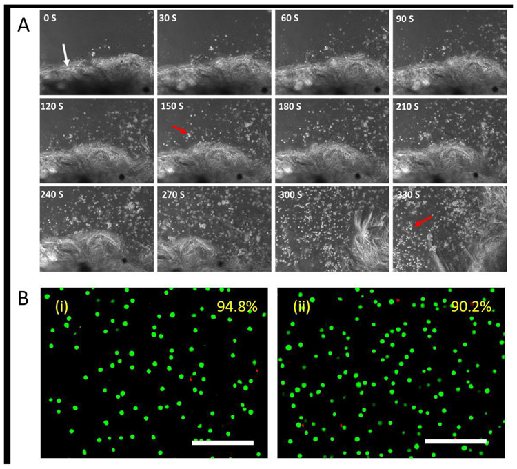
Fig. 6. Release of encapsulated cells from hydrogels upon light exposure.
A. L929 murine fibroblasts were encapsulated in SELP-CarHc hydrogels (gel boundary is indicated by white arrow) and cultured for 24 h. Gel was exposed to the white light of 30klux and to initiate cell release along with the dissociation of the hydrogel. Micrograph of light exposure and cell release is indicated at every 30 seconds. The red arrow indicates released cells from the hydrogel. B. Live/dead staining micrographs of (i) untreated control and (ii) encapsulated cells 12 h after release. Average cell viabilities estimated from 5 random images are shown in the upper right corner. Scale bars: 200 μm.

