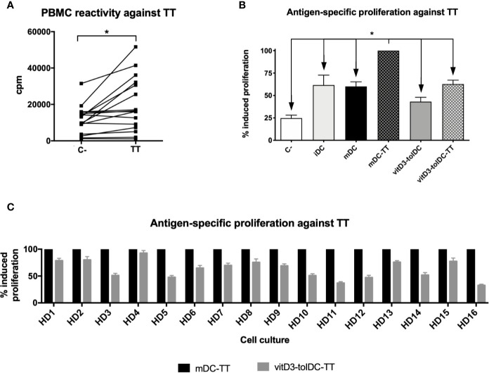
Figure 1.
PBMC reactivity and antigen-specific induction of autologous proliferation mediated by DC against tetanus toxin. (A) Induction of proliferation of PBMC without stimuli (C−) and against tetanus toxin (TT) after 5 days of culture (n = 16). Data presented as counts per minute (cpm), measured as tritiated thymidine incorporation after 18 h. Ten replicated measurements of each condition were performed. (B) Induction of antigen-specific autologous proliferation against TT mediated by immature DC (iDC), mature DC (mDC), TT-loaded mDC (mDC-TT), vitamin D3-induced tolerogenic DC (vitD3-tolDC) and TT-loaded vitD3-tolDC (vitD3-tolDC-TT), as well as a negative control (C−), without any stimuli (n = 16) and (C) comparison of autologous antigen-specific proliferation against TT mediated by mDC-TT and vitD3-tolDC-TT on each donor. Data presented as relative percentage of induced proliferation compared to mDC-TT, measured as tritiated thymidine incorporation after 18 h. Six replicated measurements of each condition were performed. Error bars corresponding to SEM. ns, not significant; *p < 0.05. One-way ANOVA test with Geisser–Greenhouse correction or paired t test.

