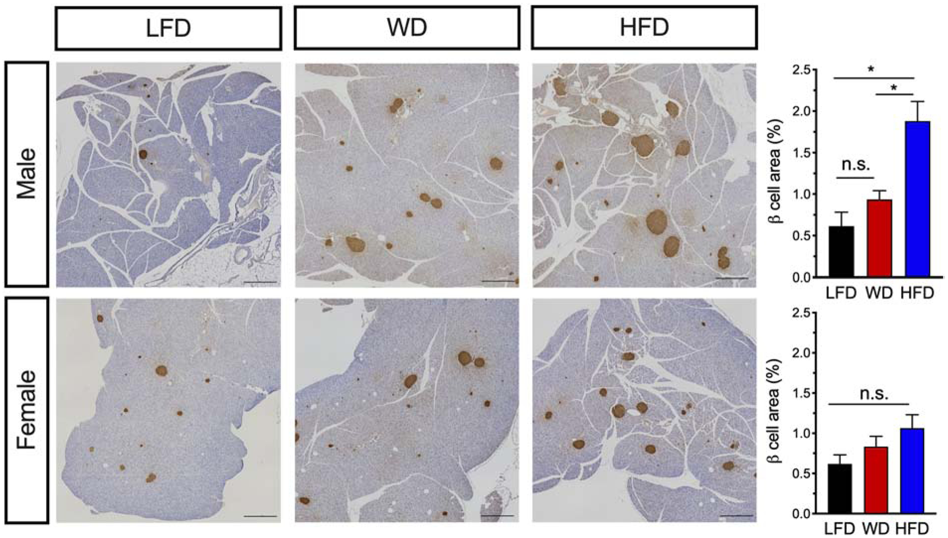
Figure 6: Male mice demonstrate increased β-cell area in response to either a WD or HFD.

Male and female mice C57BL6/J mice were placed on a dietary intervention starting at 4 weeks of age. After 22 weeks of diet, mice were euthanized and pancreata harvested and stained for insulin. Left panel: Images of whole pancreatic sections from representative mice immunostained for insulin (brown) and counterstained with hematoxylin. Scale bars, 1000 μm. Right panel: Quantitation of pancreatic β-cell area in male mice (top) and in female mice (bottom); Results are displayed as means ± SEM; n=3–5 mice per treatment group. Means were compared by one-way ANOVA; *, p<0.05 for indicated comparisons.
