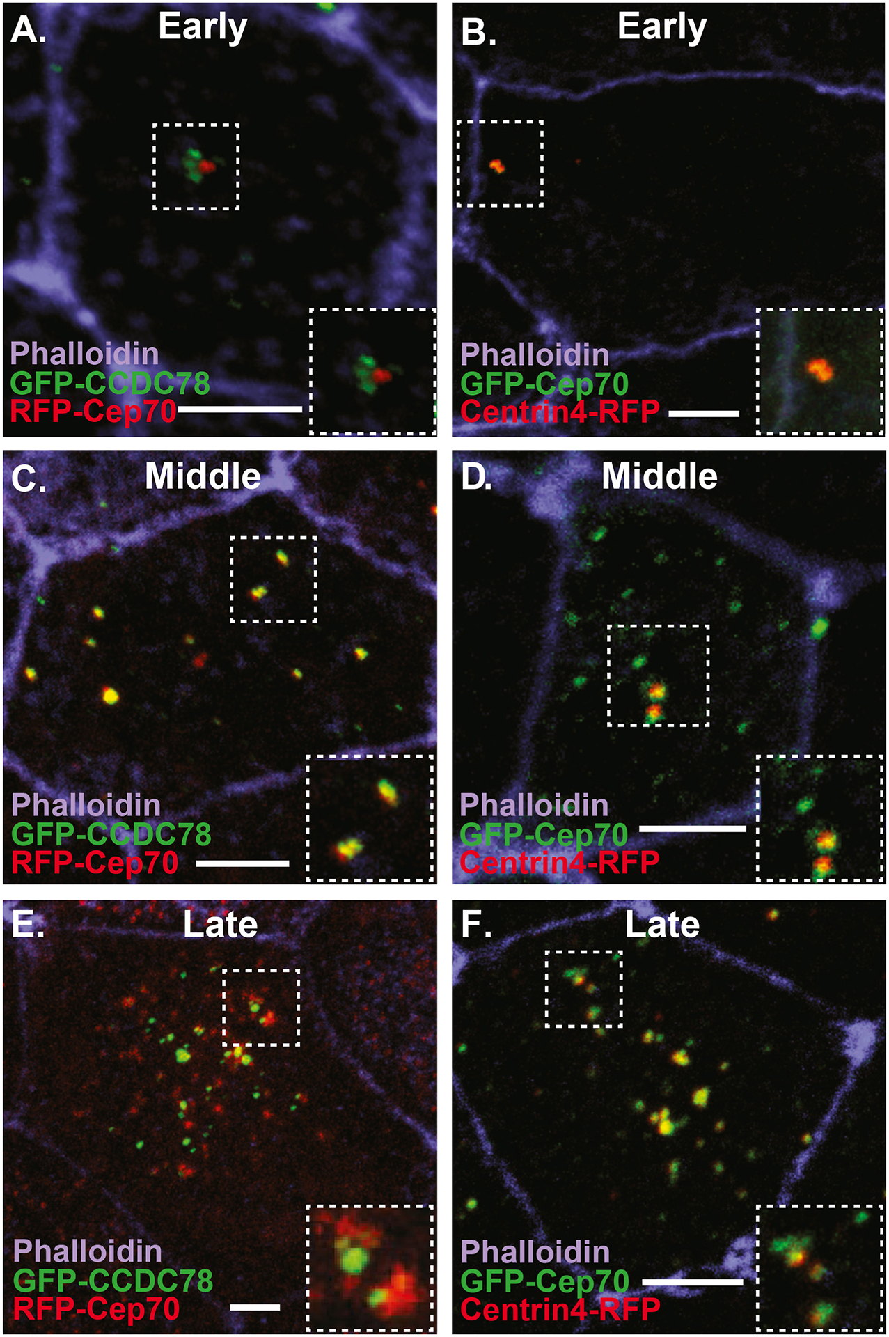
Figure 2. Dynamic localization of Cep70 to the deuterosome.

A–F Images of ectopic MCCs from embryos injected with the MCC inducing factor hGR-MCIDAS, treated with Dex and co-injected with RFP-Cep70 and the deuterosome marker GFP-CCDC78 (A, C, E) or GFP-Cep70 and the centriole marker Centrin4-RFP (B, D, F) stained with phalloidin. A–B, Early phase (2–3Hrs) of MCC development with Cep70 co-localizing to parental centrioles (B) but not nascent deuterosomes marked by CCDC78 (A). C–D, Middle phase (4–6Hrs) of MCC development showing FP-Cep70 co-localizing with GFP-CCDC78 at deuterosomes (yellow foci in E) but not at parental centrioles (red foci in C) and co-localizing with centrin4-RFP at parental centrioles (yellow foci in D) but not deuterosomes (green foci in D). Late phase (> 6Hrs) of MCC development showing FP-Cep70 co-localizing with GFP-CCDC78 at deuterosomes (yellow foci in E) but not at presumptive nascent centrioles (red foci in E) and co-localizing with Centrin4-RFP at nascent centrioles (yellow foci in F) but not presumptive deuterosomes (green foci on F). Scale bar is 5μm.
