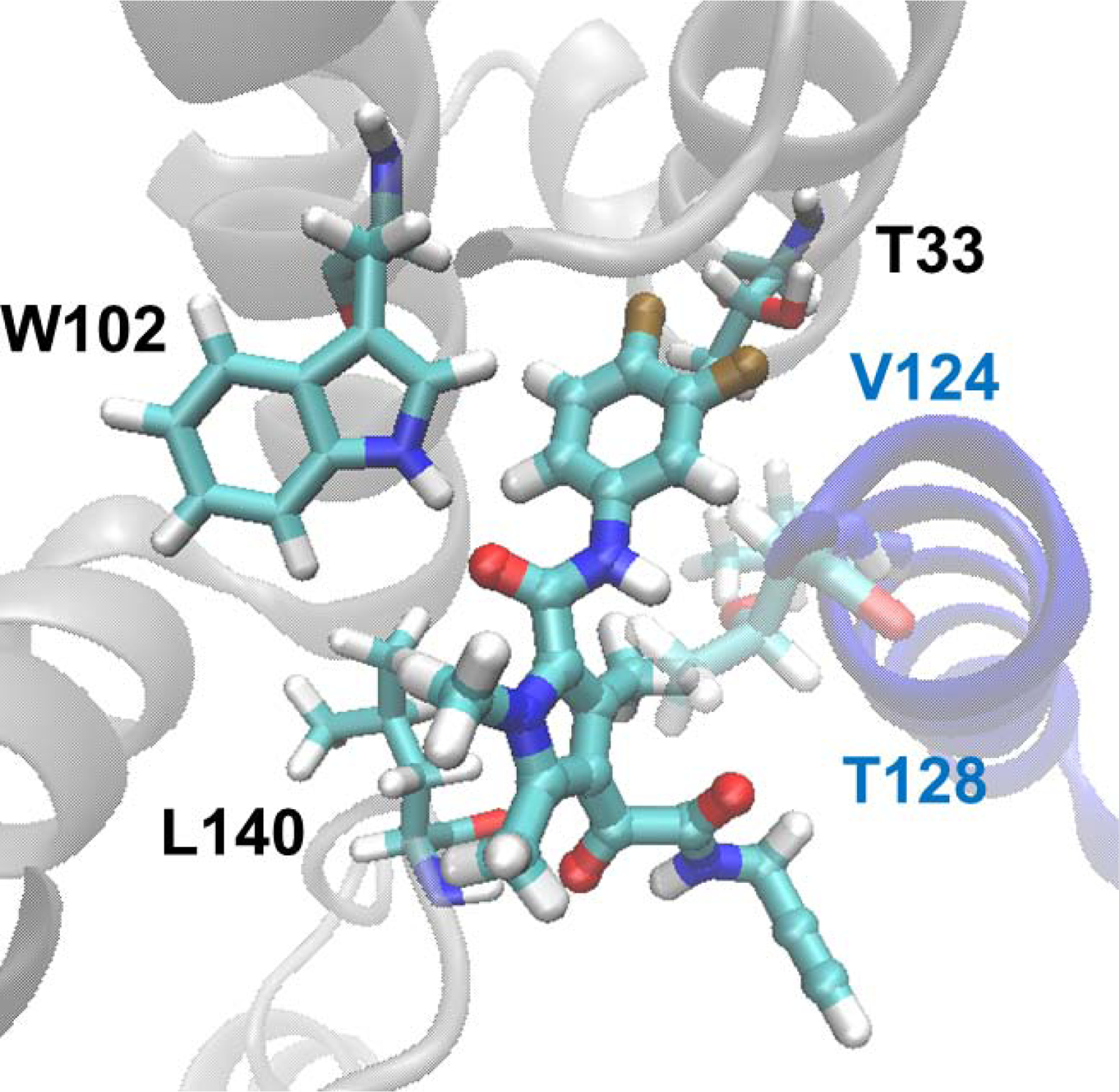
Figure 4.

Molecular model of GLP-26 bound to the HBV capsid protein interface. GLP-26 was docked into the interfacial binding pocket of compound 4 in the reported crystal structure (PDBID 5T2P) using Glide. Chain B of the protein is grey, and chain C is colored blue. Key residues are shown as licorice, and the GLP-26 is rendered as a ball-and-stick.
