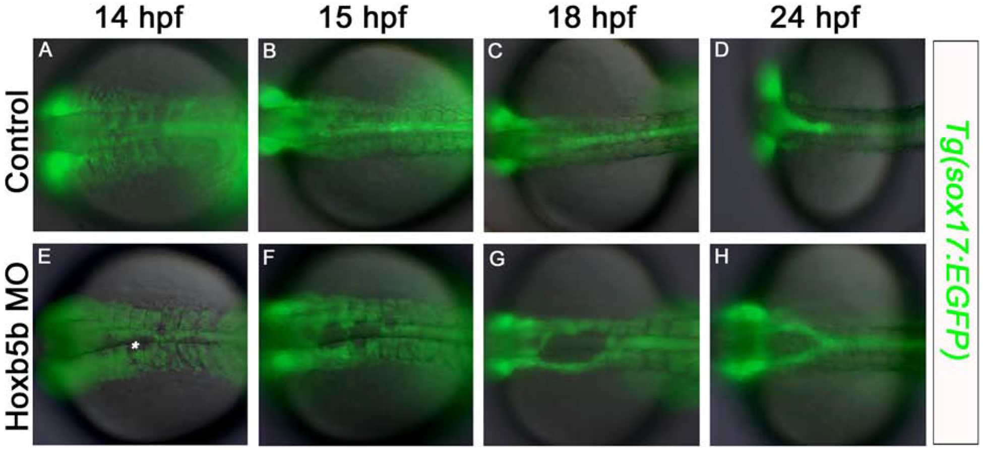
Fig. 5. Midline defects in the foregut endoderm first become apparent at 14 hpf in Hoxb5b morphants.

Fluorescent imaging of live Tg(sox17:EGFP) embryos (A-D) control, and (E-H) Hoxb5b morphant, at (A,E) 14 hpf; (B,F) 15 hpf; (C,G) 18 hpf; (D,H) 24 hpf. At 14 hpf endoderm cell-free patches are apparent at the midline in Hoxb5b-deficent specimens (asterisk). Anterior to the left, results are from 2 independent experiments and minimum of 8 embryos per group.
