Figure 3.
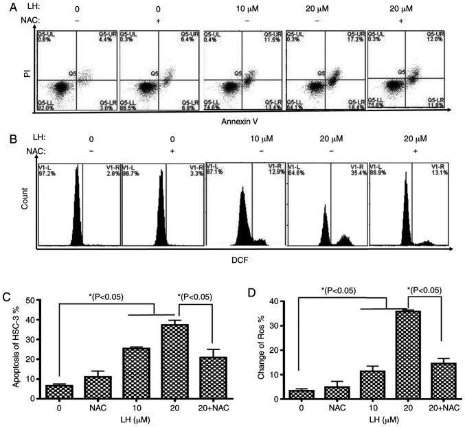
LH induces apoptosis mediated by ROS in HSC-3 cells. (A) Apoptotic effects of LH on HSC-3 cells were determined via dual staining with Annexin V-FITC and PI. Alive cells are Annexin V−/PI− (Q1-LL), cells in early apoptosis are Annexin V+/PI− (Q1-LR), cells in late apoptosis are Annexin V+/PI+ (Q1-UR) and necrotic cells are Annexin V−/PI+ (Q1-UL). (B) Intracellular ROS levels were investigated using the ROS-detecting fluorescence dye, DCFH-DA and flow cytometric analysis. (C) Population of apoptotic cells (Q1-LR and Q1-UR) was evaluated using histogram analyses. (D) Changes in ROS were investigated via histogram analyses. *P<0.05 was considered to indicate a statistically significant difference using Dunnett's test for multiple group comparisons with the control group (0 µM LH) and Tukey's test for comparisons between group differences (groups, 20 and 20 + NAC). LH, Lycorine hydrochloride; ROS, reactive oxygen species.

