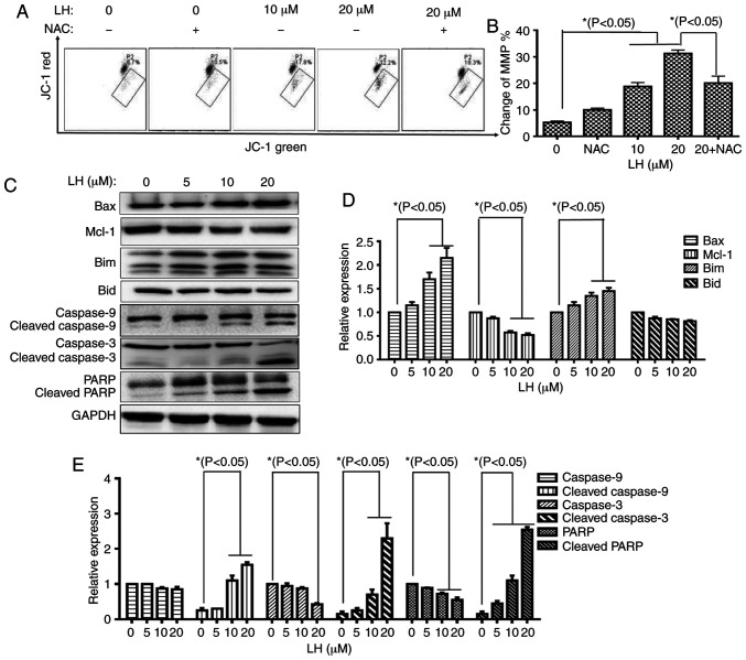
Figure 4.
LH induces HSC-3 cell apoptosis via the mitochondrial pathway. (A) Changes in MMP in HSC-3 cells were detected via JC-1. Data were analyzed using Accuri C6 FCM software by measuring green (530±30 nm) and red (585±40 nm) JC-1 fluorescence. MMP loss was observed by a decrease in JC-1 red fluorescence and an increase in JC-1 green fluorescence. In total, ≥5,000 cells were collected and counted per sample. (B) Changes in the MMP in HSC-3 cells were investigated via histogram analyses. *P<0.05 was considered to indicate a statistically significant difference using Dunnett's test for multiple group comparisons with the control group (0 µM LH) and Tukey's test for comparisons between group differences (groups, 20 and 20 + NAC). (C) Expression levels of the mitochondrial pathway-related apoptotic proteins were detected via western blot analysis. (D and E) HSC-3 cells were treated with LH for 24 h, harvested and total protein lysate was subjected to western blot analysis using antibodies against GAPDH, Bax, Bim, Mcl-1, Bid, caspase-9, caspase-3 and PARP. The apoptotic protein expression was investigated via histogram analyses. *P<0.05 was considered to indicate a statistically significant difference using Dunnett's test for multiple group comparisons with the control group (0 µM LH). LH, lycorine hydrochloride; PARP, poly(ADP-ribose) polymerase 1; Mcl-1, MCL1 apoptosis regulator, BCL-2 family member; MMP, mitochondrial membrane potential.

