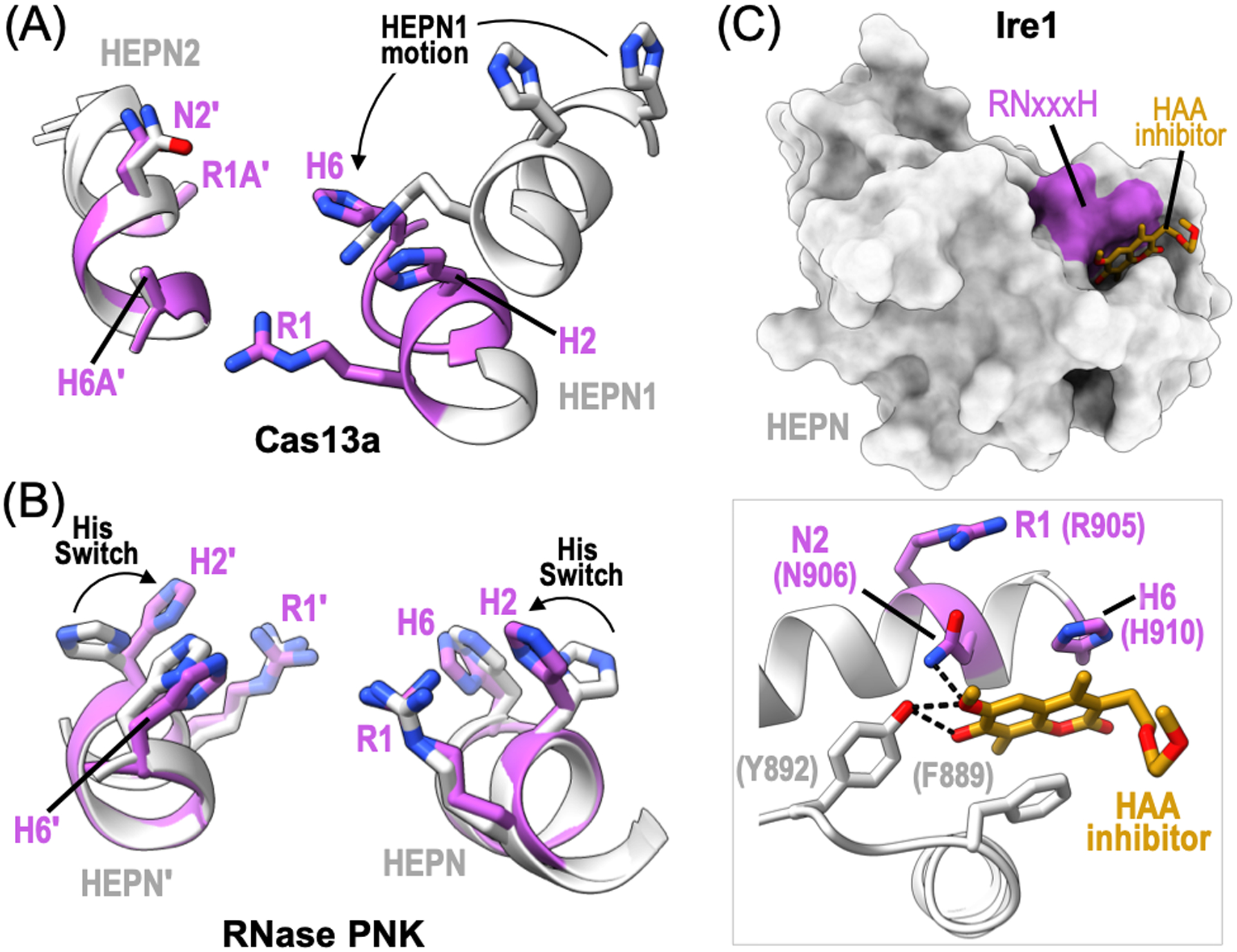
Figure 6.

HEPN RNase active site regulation. (A) Ribbon diagram of the RNase active site from L. buccalis Cas13a bound to crRNA and target RNA (PDB ID: 5XWP, 5XWY). The inactive state is shown in light grey and the active state is colored magenta. HEPN1 undergoes conformational changes, such as rearrangement of its RHxxxH motif (black arrow), for RNase activation. (B) Ribbon diagram of C. thermophilum RNase PNK RNase active site (PDB ID: 6OF2, 6OF3). The polar residue (H2) from the RNase RHxxxH motif undergoes a conformational change to orient H2 towards the catalytic center. This conserved histidine is essential for rRNA processing and is referred to as the Histidine Switch (His Switch). (C) Surface rendering of the Mus musculus Ire1 HEPN domain bound to the HAA inhibitor, MKC9989 (orange) (PDB ID: 4PL3). Inset shows a zoom in view of the HAA inhibitor binding site. Ire1 HEPN residues, such as RNase N2 and H6 residues, along with surrounding aromatic residues (grey) directly coordinate the HAA inhibitor. Dotted black lines show hydrogen bonds.
