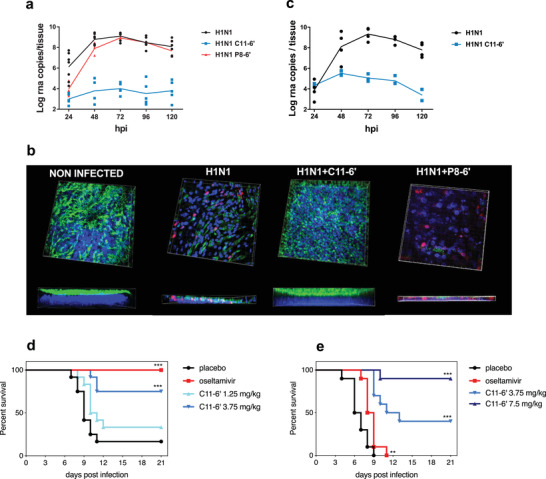
Figure 3.

Ex vivo (a to c) and in vivo (d to e) inhibitory activity of C11‐6’. a) Ex vivo, C11‐6’ provided full protection against clinical H1N1 pandemic 09 strain in co‐treatment condition, whereas P8‐6’ only provided minor protection at the beginning of the infection. b) Immunofluorescence at 7 days post‐infection (co‐treatment condition) confirms the protection provided by C11‐6’. Red: monoclonal antibody influenza A, blue: DAPI, green: β‐IV‐tubulin (marker of ciliated cells). The thickness of each tissue is shown at the bottom of the corresponding image. c) C11‐6’ also showed high efficacy in post‐treatment condition. Results of (a) and (c) are mean and SEM of 2 to 4 independent experiments with intra‐experimental duplicates. Images of (b) are representative of 10 images taken for each condition. In vivo, mice (12/group) were intra‐nasally treated with PBS or C11‐6’ or by oral gavage with oseltamivir and infected with A/California/09. d) Subsequent treatments were administered daily for the following 5 days, image shows the survival curve. e) Mice were infected and treated with C11‐6’ or oseltamivir at 24 hpi and daily for the 4 following days, images show the survival curves ***p<0.001, **p<0.01
