
Figure 6.
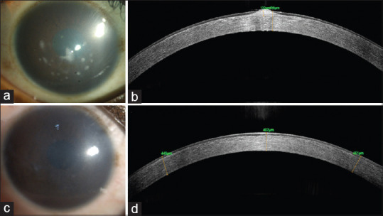
A 18-year-old male presented with photophobia and decrease in vision. (a) Diffuse slit-lamp photograph right cornea showing a diffuse corneal haze with elevated nodular deposits in the central cornea. (b) Corresponding AS-OCT of the right eye cornea showing focally elevated lesions. (c) Diffuse slit-lamp photograph of the right cornea after PTK demonstrating removal of the nodular deposits. (d) Corresponding post-PTK AS-OCT of the same area showing a slight depression that has been smoothened by epithelium