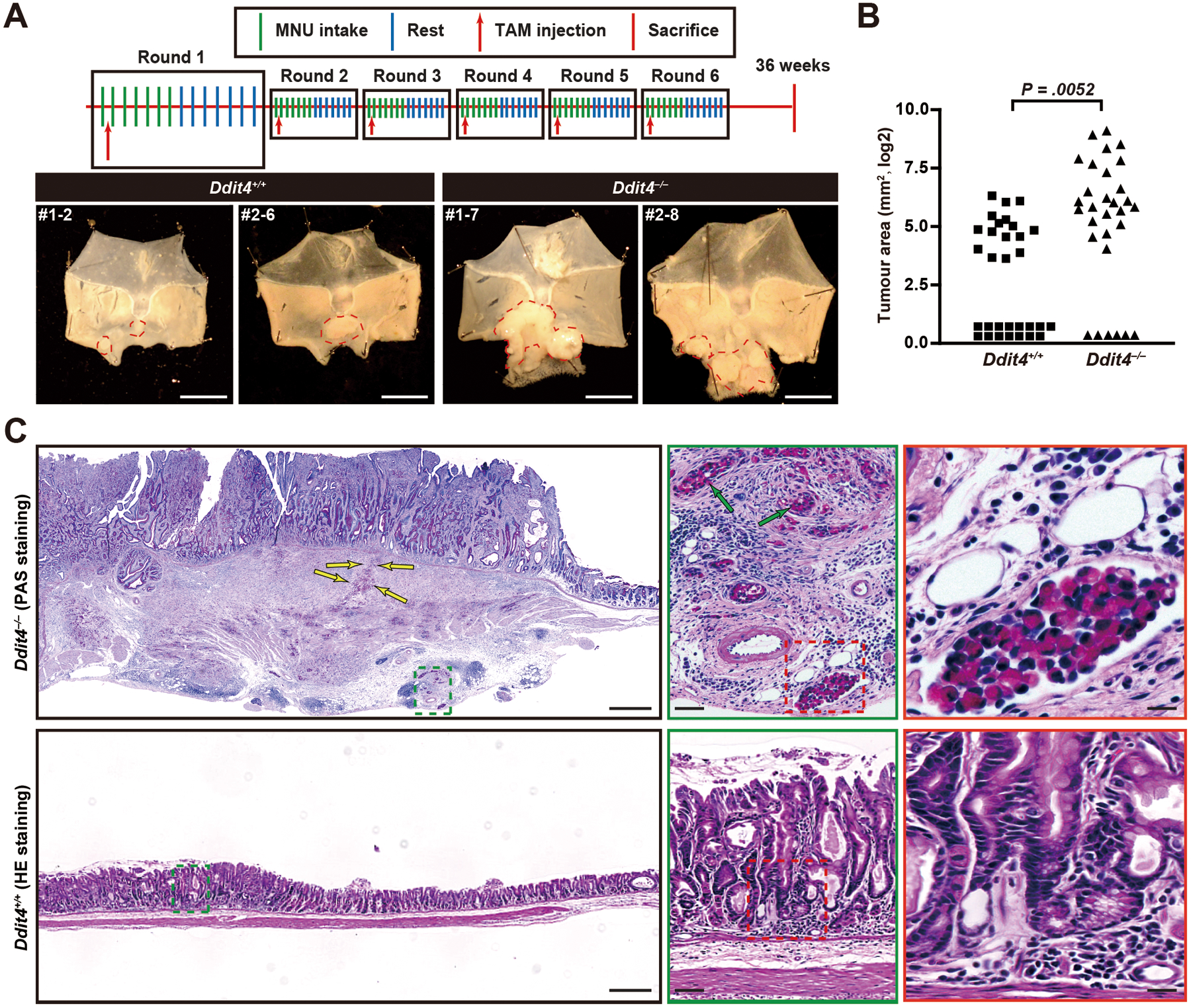
Figure 6. Loss of DDIT4 increases spontaneous tumorigenesis after multiple rounds of paligenosis in a mouse model.

A. Scheme for TAM and MNU administration (top) and example tumors (bottom) in stomachs from 40 mice per genotype (30 Ddit4−/−, 31 Ddit4+/+ mice survived until endpoint of observation) in three independent cohorts. First number is cohort #, second is individual mouse number from that cohort. Tumor area outlined by red dashed line.
B. Total area per mouse occupied by tumors as measured under dissecting microscope from stomachs opened as for (B) quantified from all mice surviving until the endpoint of observation. Each datapoint: area of single mouse tumor area, log2 scale; significance estimated by Mann-Whitney test.
C. Representative H&E and PAS (for mucins) of one tumor from each mouse genotype. Tumors in wildtype mice were all raised, hyperplastic lesions with focal cellular and glandular atypia but no invasive features (red and green boxes show increased magnification). In the Ddit4−/− tumor depicted (1 of 6 invasive tumors, #1–7), a massive, poorly differentiated tumor with both irregular glandular and signet ring (PAS-staining positive) morphologies is seen invading through muscle to serosa. Invasive signet ring cells are marked by yellow arrows. Scale bar, 500 μm; green arrows = perineural infiltration, scale bar 50 μm; red box shows lymph-vascular space invasion, 25 μm.
