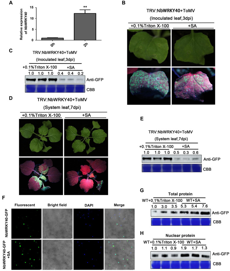
FIGURE 6.
Effect of exogenous salicylic acid (SA) application on viral infection and subcellular localization of NbWRKY40. (A) Relative expression of NbWRKY40 in response to SA treatment (B) GFP fluorescence of NbWRKY40-silenced leaves inoculated with ToMV-GFP at 3 days postinfiltration (dpi) following SA treatment is less intense than that following 0.1% Triton X-100 treatment alone. (C) Western blot analysis of GFP accumulation in ToMV-GFP-inoculated leaves at 3 dpi. Coomassie brilliant blue (CBB)-stained rubisco gel and ImageJ (United States National Institutes of Health, http://rsb.info.nih.gov/nih-image/) were used to determine protein loadings. (D) GFP fluorescence of systemic leaves of NbWRKY40-silenced plants inoculated with ToMV-GFP at 7 dpi following SA treatment is less intense than that following 0.1% Triton X-100 treatment alone. (E) Western blot analysis of GFP accumulation in non-infected leaves of ToMV-GFP-inoculated plants at 7 dpi. CBB-stained rubisco gel and ImageJ (United States National Institutes of Health, http://rsb.info.nih.gov/nih-image/) were used to determine protein loadings. (F) Subcellular localization of NbWRKY40 protein in response to SA treatment. The recombinant plasmid NbWRKY40-GFP was introduced into Nicotiana benthamiana epidermal cells using Agrobacterium infiltration. Leaves infiltrated with an Agrobacterium culture that only contained GFP acted as controls. The confocal microscopy images were captured under bright-field fluorescence to show cell morphology; under dark field to show green fluorescence, indicating localization of the NbWRKY40 protein, and blue fluorescence, indicating nuclei stained blue by 4,6-diamidino-2-phenyl-indole dihydrochloride (DAPI); and under combination fluorescence to show the three images merged. Scale bar, 100 μm. (G) Immunoblot of total protein extracted from the leaves of SA- and mock-treated plants. Anti-GFP was used to detect GFP fractions. Equal amounts of protein were used for immunoblotting and for staining with CBB. (H) Immunoblot of nuclear protein extracted from the leaves of SA- and mock-treated plants. Anti-GFP was used to detect GFP fractions. Equal amounts of protein were used for immunoblotting and for staining with CBB. **P < 0.01.

