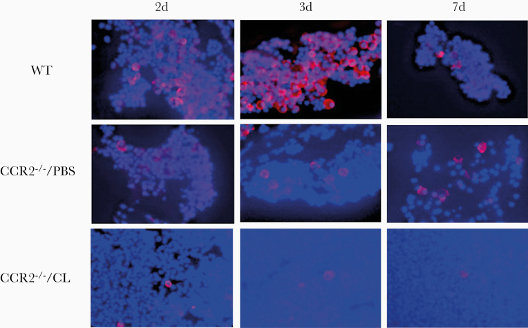
Figure 2.
Immunohistochemistry of macrophages in middle ear (ME) effusions. The macrophage marker AIF1 (allograft inflammatory factor 1) is labeled with rhodamine, and cell nuclei are labeled with DAPI. AIF1-negative cells are primarily neutrophils. The decrease in ME macrophages for CCR2−/−/PBS mice is readily apparent. The figure also illustrates the scarcity of macrophages, as well as the persistence of substantial cellular infiltrate at 7 days (7d) after NTHi inoculation, in the CCR2−/−/CL MEs.

