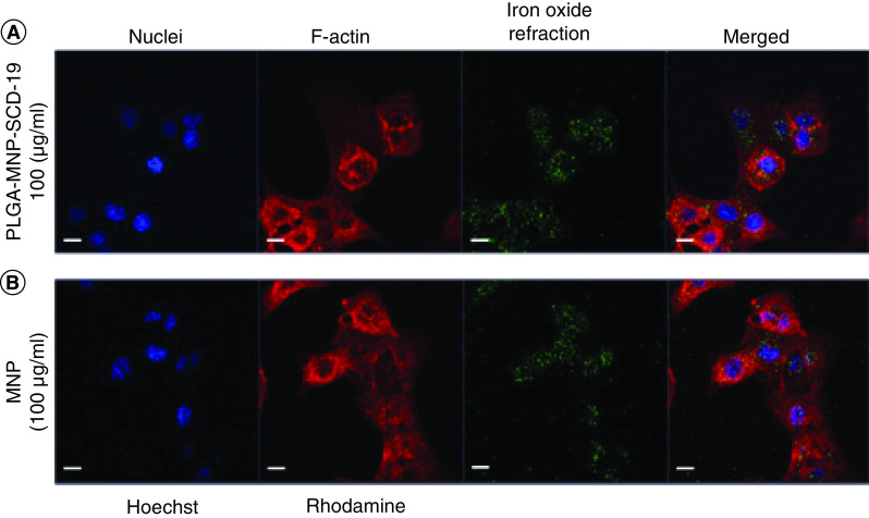
Figure 2. . Internalization of poly lactic-co-glycolic acid-magnetic nanoparticles-SCD-19 nanoparticles by A549 cells.
The confocal images were acquired after cell exposure topoly lactic-co-glycolic acid-magnetic nanoparticles-SCD-19 and uncoated magnetic nanoparticles for 24 h. The cells were stained with Hoechst 33342 and Rhodamine phalloidin to stain nuclei (blue) and actin filaments (red), respectively. (A) The A549 cells exposed to PLGA-MNP-SCD-19 (100 μg/ml). (B) The positive control was only treated by uncoated magnetic nanoparticles (100 μg/ml). Scale bar 10 μm.
MNP: Magnetic nanoparticle; NP: nanoparticle; PLGA: Poly lactic-co-glycolic acid.

