Figure 1.

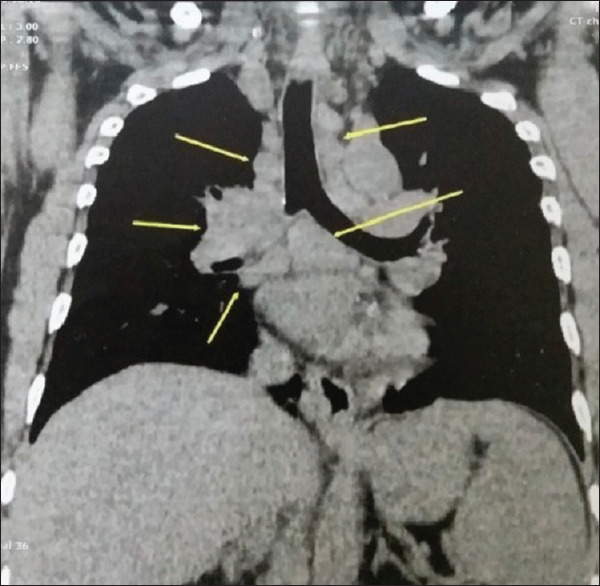
Computed tomography thorax sagittal image, mediastinal window showing enlarged paratracheal, subcarinal, and hilar lymph nodes (arrows)

Computed tomography thorax sagittal image, mediastinal window showing enlarged paratracheal, subcarinal, and hilar lymph nodes (arrows)