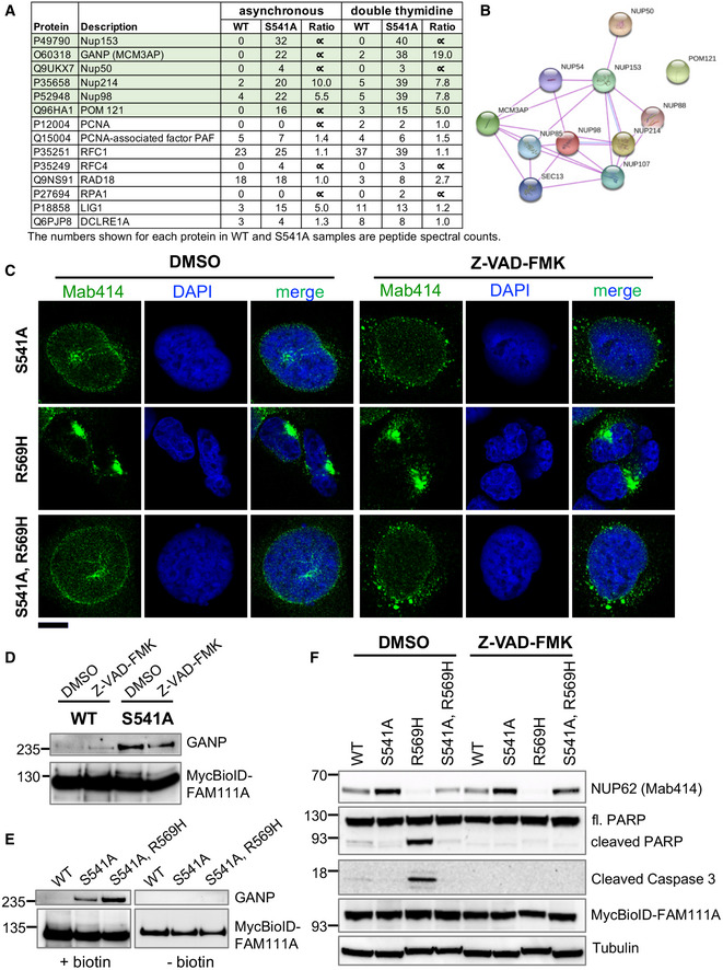
A subset of FAM111A interacting proteins identified by BioID screen is listed along with the number of peptide spectral counts observed for each protein.
A STRING diagram showing the interaction of NPC components identified in our FAM111A BioID screen.
Cells were treated with doxycycline along with DMSO or Z‐VAD‐FMK for 24 h. They were fixed with formaldehyde and stained with NPC antibody Mab414 and DAPI. The images were captured by confocal microscopy. Scale bar: 10 μm.
DMSO or Z‐VAD‐FMK‐treated HEK293/MycBioID‐FAM111A cell lines were induced with doxycycline for 24 h. Total protein from an equal number of cells was incubated with magnetic streptavidin beads, and pull downs were analyzed by Western blotting using GANP and Myc antibodies.
HEK293/MycBioID‐FAM111A cell lines were induced with doxycycline for 24 h, while growing in the presence or absence of 50 µM supplemental biotin. Total protein from whole cell lysates was incubated with magnetic streptavidin beads. Pulldowns were analyzed by Western blotting using GANP and Myc antibodies.
DMSO or Z‐VAD‐FMK‐treated HEK293/MycBioID‐FAM111A cell lines were induced with doxycycline for 24 h. Total protein from ~105 cells was resolved by SDS–PAGE and probed in Western blots using antibodies against NPC (Mab414), PARP, cleaved Caspase 3, Myc, or Tubulin.

