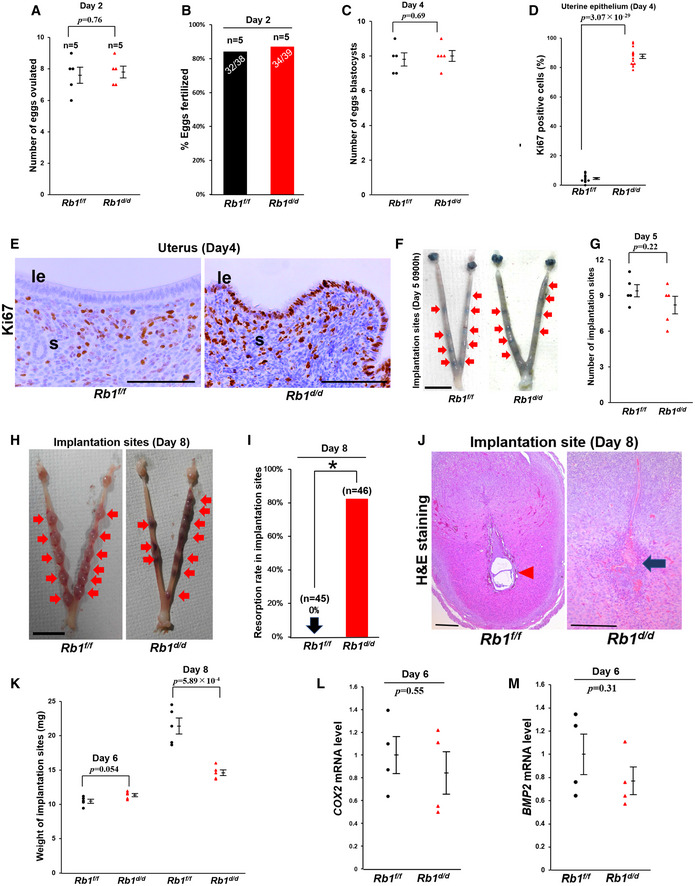
Figure 2. Rb1d/d mice show impaired pre‐implantation epithelial cell cycle arrest and implantation failure.

-
A–COvulation, fertilization, and development of pre‐implantation embryos were normal in Rb1d/d mice (A and C, mean ± SEM, Student’s t‐test; n = 5 mice for each group; B, Fisher's exact test; n = 5 mice for each group).
-
D, ENumber of Ki67‐positive cells on day 4 in uterine epithelium of Rb1d/d mice was higher than that of Rb1f/f mice (mean ± SEM, Student’s t‐test; n = 5 mice for each group). Scale bar = 100 μm; le, luminal epithelium; s, stroma.
-
F, GEmbryo attachment occurred normally in Rb1d/d mice at 09:00 h on day 5 (mean ± SEM, Student’s t‐test; n = 5 mice for each group). Arrow, implantation site; scale bar = 1 cm.
-
H, IRb1d/d mice showed higher rate of embryo resorption on day 8 (*P < 0.05; Fisher's exact test; n = 45 implantation sites for Rb1f/f mice and n = 46 implantation sites for Rb1d/d mice). Arrow, implantation site; scale bar = 1 cm.
-
JH&E staining showed embryo resorption in Rb1d/d mice on day 8 of pregnancy. Scale bar = 200 μm; arrowhead, embryo; arrow, broken embryo with blood cell infiltration.
-
KWeight of implantation site was comparable between Rb1f/f and Rb1d/d mice on day 6, but reduced on day 8 in Rb1d/d mice compared with Rb1f/f mice (mean ± SEM, Student’s t‐test; n = 6 mice for each group).
-
L, MExpression of Cox2 and Bmp2, markers of decidualization, was comparable in between Rb1d/d and Rb1f/f mice at 09:00 h on day 6 in implantation sites (mean ± SEM, Student’s t‐test; n = 4 mice for each group).
Data information: le, luminal epithelium; s, stroma; tr, trophoblast; n, nucleus.
