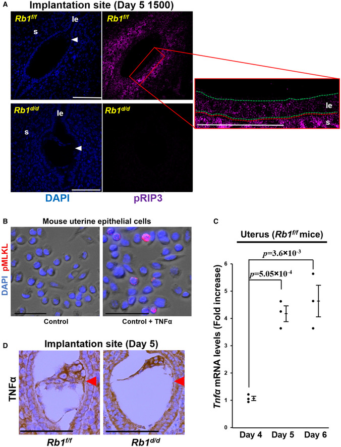
pRIP3, a central mediator of necroptosis, was not expressed at the implantation sites of Rb1d/d mice but was expressed at those of Rb1f/f mice at 15:00 h on day 5. Scale bar = 100 μm; blue signal, nuclei stained by DAPI; purple signal, pRIP3; le, luminal epithelium; s, stroma; arrowhead, embryo; green dotted line, luminal epithelium; red dotted line, stroma.
TNFα stimulated the expression of pMLKL, a critical mediator of necroptosis, in the primary mouse uterine epithelial cells. Scale bar = 50 μm; blue signal, nuclei stained by DAPI; red signal, pMLKL.
Uterine expression of TNFα was elevated in the control mice (Rb1f/f mice) on days 5 and 6 compared with on day 4 (mean ± SEM, Student’s t‐test; n = 3 mice for each group). As for the uterine samples on days 5 and 6, those with implantation sites were examined.
The implanting embryo and the uterus produced TNFα at the implantation site of both Rb1f/f and Rb1d/d mice. Immunostaining of TNFα at the implantation sites of Rb1f/f and Rb1d/d mice were performed. Scale bar = 100 μm; arrowhead, embryo.