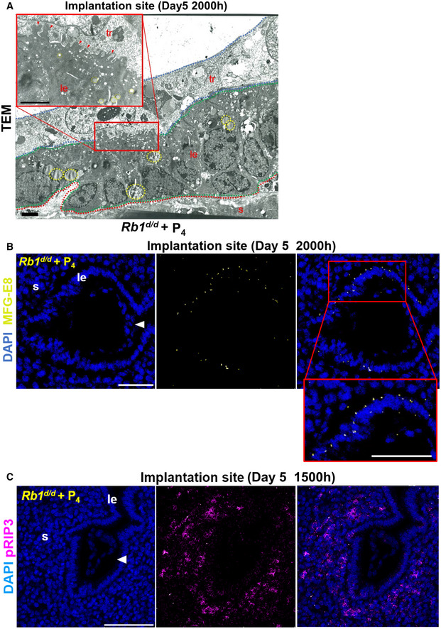
In Rb1d/d mice with pre‐implantation P4 treatment, the fragmented uterine epithelial cells with the cytoplasmic lipid droplets were engulfed by the trophoblast cells. Scale bar = 2 µm. Arrowhead, cytoplasmic fragments engulfed by trophoblast; dotted line circle, lipid droplets in the cytoplasm; tr, trophoblast; le, luminal epithelium; s, stroma; red dotted line, stroma; green dotted line, luminal epithelium; blue dotted line, trophoblast.
Pre‐implantation P4 treatment rescued the expression of MFG‐E8 on day 5 of pregnancy in Rb1d/d uteri. Scale bar = 100 μm. Blue signal, nuclei stained by DAPI; yellow signal, MFG‐E8; le, luminal epithelium; s, stroma; arrowhead, embryo.
Pre‐implantation P4 treatment recovered the expression of pRIP3, a mediator of necroptosis, on day 5 of pregnancy in Rb1d/d uteri. Scale bar = 100 μm; blue signal, nuclei stained by DAPI; purple signal, pRIP3; le, luminal epithelium; s, stroma; arrowhead, embryo.