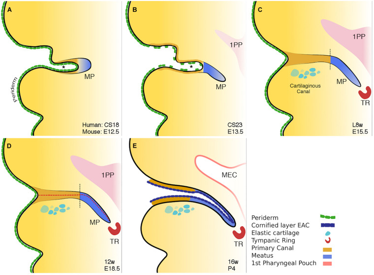
FIGURE 3.
Schematic of mammalian ear canal development. (A) Invagination of the primary canal (marked by *) with a meatal plug at its tip. (B) Extension of the meatus toward the forming middle ear, driven by a potential signal from the tympanic ring. At the same time selective loss of periderm is observed in the primary canal caused by apoptosis. (C) Closure of the primary canal (following loss of periderm layer) and further extension of the meatal plug to reach the tympanic ring. (D) Loricrin expression (red dashed line) marks the site of opening of the canal. (E) Following keratinocyte differentiation, the whole external ear canal opens, with the upper/rostral wall of the meatal plate forming the outer surface of the tympanic membrane. Vertical black dashed lines in C and D between brown and blue regions represent the two distinct parts of the ear canal. Schematic taken from Fons et al. (2020).

