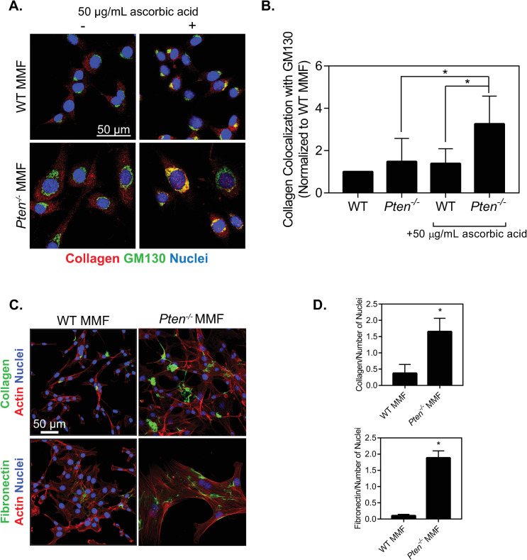
Fig 3. Pten knockout alters collagen shuttling and fibrillogenesis.
(A) Immunofluorescence images of WT and Pten-/- MMF plated for 24 hours and cultured with or without 50 μg/mL ascorbic acid for 1 hour, then fixed and stained for collagen, GM130, and nuclei. (B) Quantification of collagen colocalization with GM130, determined by Manders’ correlation coefficient and normalized to the WT condition. n = 5+SD. *p<0.05. (C) Immunofluorescence images of cells incubated with fluorescently labeled collagen and fibronectin for 24 hours, then fixed and stained for actin and nuclei. (D) Quantification of the area of each image covered by collagen fibers normalized to number of nuclei in each image. (E) Quantification of the area of each image covered by fibronectin fibers normalized to number of nuclei in each image. n = 3+SD. *p<0.05 relative to WT.

