Figure 1.

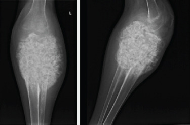
AP and lateral X-rays of the left knee demonstrating a large mineralized mass encompassing the proximal 20 cm of the tibia and fibula with obliteration of the joint and extension into the surrounding soft tissues.

AP and lateral X-rays of the left knee demonstrating a large mineralized mass encompassing the proximal 20 cm of the tibia and fibula with obliteration of the joint and extension into the surrounding soft tissues.