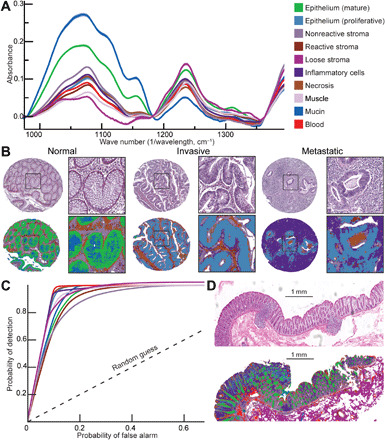
Fig. 2. Pixel-level precise segmentation of tissue components using IR spectroscopic imaging.

(A) Intrinsic chemical contrast in tissue produces unique IR spectrum for each of the 10 histologic classes. (B) Performance of the artificial intelligence classifier in segmenting normal and invasive tumor containing lymph node tissue cores. (C) ROC curve demonstrating high performance of artificially intelligent classifier in segmenting tissue into 10 unique components as compared with an experienced pathologist. (D) Demonstration of segmentation accuracy on a surgical resection. The color key is common across panels.
