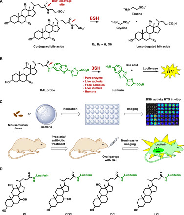
Fig. 1. Design and applications of BAL probes.

(A) Enzymatic deconjugation mechanism of naturally occurring conjugated BAs. (B) Schematic representation of the BAL assay. The technology is based on BSH-specific uncaging of free luciferin, which is subsequently processed by the luciferase enzyme to produce bioluminescent light. Hence, the signal output is proportional to the level of BSH activity. (C) The BAL method is suitable for imaging and quantification of BSH activity in different settings, including pure in vitro enzymes, cultured bacteria, and mouse and human fecal samples, as well as noninvasive real-time imaging and quantification directly in vivo in both normal and altered physiological conditions (e.g., antibiotic/probiotic treatment). The BAL probes administered to the animals expressing luciferase (FBV + luc) via oral gavage. Immediately after gavage, the animals are imaged using a CCD camera to quantify the resulting signal as a function of time. GIT, gastrointestinal tract. (D) Structures of the BAL probes used here. The design is based on four principal BAs: two primary BAs, namely, choloyl-luciferin (CL) and chenodeoxycholoyl-luciferin (CDCL); and two secondary BAs, namely, deoxycholoyl-luciferin (DCL) and lithocholoyl-luciferin (LCL).
