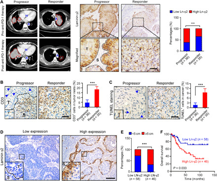
Fig. 1. High Ln-γ2 expression predicts a diminished response of patients with NSCLC to anti–PD-1 therapy.

(A) Representative images of computed tomography scans and IHC staining in patients with NSCLC treated with anti–PD-1 (nivolumab) therapy showed significantly higher Ln-γ2 expression in tumor tissues from progressors (n = 36) than in tumor tissues from responders (n = 35). In the left panel, NSCLC tumors are indicated by red dotted lines. H, heart; A, aorta; T, tumor; S, stroma. Scale bar, 100 μm. (B) IHC staining with an antibody against CD3 to detect T cells in NSCLC tissues. Blue arrows indicate CD3+ cells in the tumor. Scale bar, 100 μm. (C) Apoptotic cells in NSCLC tissues from progressors and responders were detected using TUNEL staining. Blue arrows indicate the TUNEL+ cells. Scale bar, 100 μm. (D) Representative images of IHC staining for low or high Ln-γ2 expression in NSCLC tissues. Scale bar, 200 μm. (E) The correlation analysis revealed a positive correlation between high Ln-γ2 expression and the tumor size in patients with NSCLC (total n = 104). (F) Survival curves indicated that high expression of Ln-γ2 (n = 46) predicted shorter survival of patients with NSCLC compared with patients with low expression of Ln-γ2 (n = 58). The data presented in (A) and (E) were analyzed using Pearson’s chi-square (Fisher’s exact) test, statistical analyses of the data presented in (B) and (C) were performed using unpaired two-tailed t test with Welch’s correction, and the data presented in (F) were analyzed with the log-rank (Mantel-Cox) test. In all panels, ***P < 0.001.
