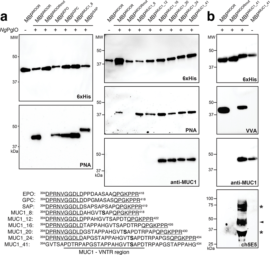
Figure 5. O-linked glycosylation of diverse protein targets.
(a) Immunoblot analysis of acceptor proteins purified from CLM25 cells co-transformed with pOG-T-NgPglO (+) or pOG-T without NgPglO (−) along with pEXT-based plasmid encoding each of the different protein targets as indicated. Absence of NgPglO or mutation of acceptor serine to glycine in MBPMOORmut served as negative controls. Blots were probed with anti-hexa-histidine antibody (6xHis) to detect acceptor proteins and PNA lectin to detect the T antigen. Additional blot for MUC1 variants was probed with murine H23 antibody (anti-MUC1) that is specific for APDTRP motif in human MUC1. Shown at bottom are acceptor sequences derived from human EPO, GPC, and MUC1 as well as synthetic SAP. All acceptor motifs except for MUC1_41 are presented in the context of the hydrophilic flanking regions derived from the MOOR tag (underline). MUC_41 was designed without hydrophilic flanking residues and includes the VNTR region as indicated. Serine amino acids determined to be glycosylated by EThcD fragmentation analysis are shown in bold font. (b) Immunoblot analysis of MUC1_41 expressed in CLM25 cells carrying pOG-Tn-NgPglO (+) or pOG-Tn without NgPglO (−). Also shown is MBPMOOR and MBPMOORmut derived from the same cells. Blots were probed with anti-6xHis antibody to detect acceptor proteins, VVA lectin to detect the Tn antigen, anti-MUC1 to detect MUC1_41, and chimeric 5E5 antibody (ch5E5) to detect Tn-MUC1. Arrow denotes the expected Tn-MUC1 glycoform, while asterisks denote higher and lower molecular weight species that may represent SDS-stable multimers and degradation products, respectively. Molecular weight (MW) markers are indicated on the left of each blot. All immunoblot results are representative of at least three biological replicates. See Supplementary Fig. 3 for uncropped versions of the images.

