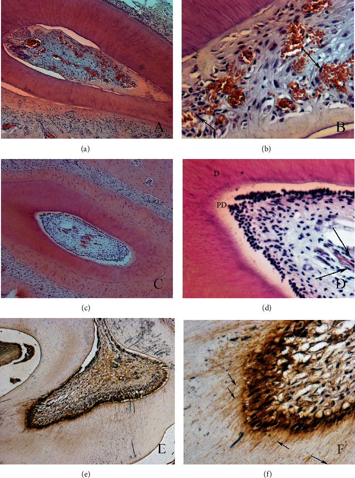
Figure 5.

Photomicrographs illustrating the results of the blood clot+chitosan+PBMT group. Observe the newly formed connective tissue inside the root canal (a, b), with young blood vessels. This newly formed tissue is similar to healthy dental pulp (c, d). The odontoblast-like cells stained with HSP-25 (e, f) are in intimate contact with the predentin (original magnifications of (e) 10x and (f) 40x).
